Human Heart Photo

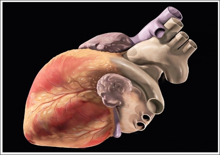
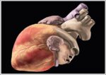
Human Heart Photo

The human heart, about fist-sized and nestled in the mediastinum behind the sternum, has a pointed apex at the left fifth intercostal space and base at the second rib, with…
Heart Valves

Heart Valves

Heart valves ensure one-way blood flow through precisely timed opening and closing. Atrioventricular valves—the tricuspid on the right and mitral on the left—hang from chordae tendineae anchored to papillary muscles,…
Types of Synovial Joint Diagram

Types of Synovial Joint Diagram

Types of synovial joints: plane (gliding, e.g., intercarpal), hinge (uniaxial, e.g., elbow), pivot (rotation, e.g., atlantoaxial), condyloid (biaxial, e.g., wrist), saddle (biaxial, e.g., thumb CMC), ball-and-socket (multiaxial, e.g., hip). All…
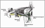
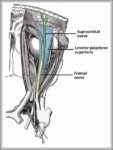
Course of the Lacrimal Nerve Diagram

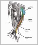
Course of the Lacrimal Nerve Diagram

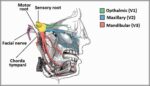
Lacrimal nerve (V1 branch) enters orbit via superior orbital fissure, runs superolateral, supplies lacrimal gland (parasympathetic hitchhike from facial via pterygopalatine ganglion), conjunctiva, upper eyelid skin. It communicates with zygomatic…
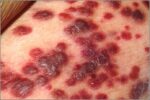
Kaposis Sacroma Lesions

Kaposis Sacroma Lesions

Kaposi's sarcoma lesions appear as purple-red plaques or nodules on skin, mucosa, or viscera, caused by HHV-8 in immunocompromised like AIDS patients, proliferating spindle cells with vascular slits leaking red…
Anatomical Relations of the Prostate Gland Diagram

Anatomical Relations of the Prostate Gland Diagram

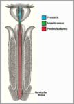
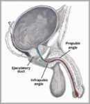
Anatomical relations of prostate gland: anterior pubic symphysis/vesicoprostatic space, posterior rectum (separated by Denonvilliers’ fascia), superior bladder neck, inferior urogenital diaphragm, lateral levator ani. Seminal vesicles posterior-superior; urethra passes through.
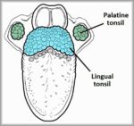
The Lingual Tonsil Waldeyers Ring Diagram

The Lingual Tonsil Waldeyers Ring Diagram

The lingual tonsil forms part of Waldeyer’s ring on posterior tongue base, composed of lymphoid follicles. It guards oropharynx entrance, enlarges in infection/allergy, visible on endoscopy or lateral neck X-ray.
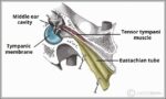
Tensor Tympani Muscle Middle Ear Diagram

Tensor Tympani Muscle Middle Ear Diagram

Tensor Tympani Muscle Middle Ear diagram shows this tiny muscle originating from the Eustachian tube cartilage and sphenoid, inserting on the malleus handle to dampen loud sounds by tensing the…
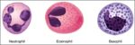
Granular Leukocytes

Granular Leukocytes

Granular leukocytes, or granulocytes, are white blood cells with distinctive cytoplasmic granules and lobed nuclei, comprising neutrophils, eosinophils, and basophils. Neutrophils, the most abundant, rush to infection sites to phagocytose…
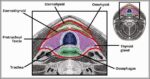
Diagram of the Pretracheal Fascia Layer of the Neck Labelled Diagram

Pretracheal Fascia Layer of the Neck Labelled Diagram

Pretracheal fascia (visceral layer) invests thyroid, trachea, esophagus, infrahyoid muscles; blends with carotid sheath laterally, buccopharyngeal fascia posteriorly. It forms “visceral vascular space” and invests recurrent laryngeal nerves.
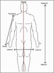
anatomical terms of location Diagram

anatomical terms of location Diagram

Anatomical terms of location standardize descriptions: superior/inferior, anterior/posterior, medial/lateral, proximal/distal, superficial/deep, palmar/plantar, dorsal/ventral, etc., based on anatomical position.