
ECG tracing with heart contraction synchronizes electrical and mechanical events, where the P wave reflects atrial depolarization initiating contraction to fill ventricles, followed by the flat PR interval during AV delay, then the sharp QRS complex as ventricles depolarize and contract powerfully to eject blood, and the T wave during repolarization as ventricles relax for refilling. Isovolumetric phases align with closed valves, systole with QRS to T, diastole after, all on a timed scale where intervals like QT indicate risks if prolonged; this correlation allows diagnosis of dyssynchrony or blocks by comparing electrical signals to expected mechanical outcomes.
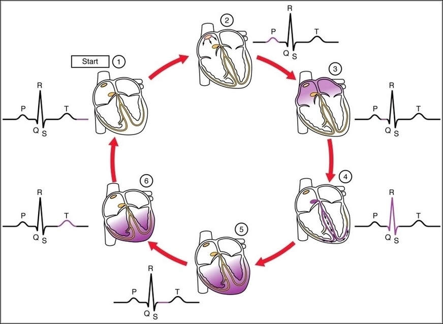
ECG Tracing with Heart ContractionN
Posted inMedical
ECG Tracing with Heart ContractionN
Post navigation
Previous Post
 Borders of the Femoral Canal Diagram
