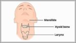
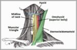
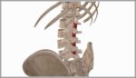
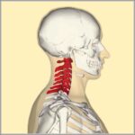
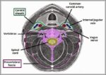
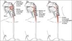
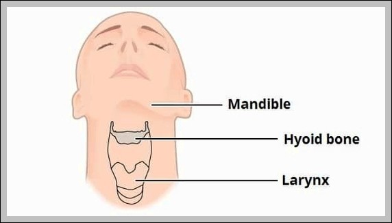
Anatomical Position and Relations of Pituitary Gland Diagram

Anatomical Position and Relations of Pituitary Gland Diagram

The pituitary gland sits in sella turcica, attached to hypothalamus by infundibulum, surrounded by cavernous sinuses laterally, optic chiasm superiorly, sphenoid sinus inferiorly. Relations affect surgical/transsphenoidal approaches and compression symptoms.
Regional Blood Supply to the Cerebrum Diagram

Regional Blood Supply to the Cerebrum Diagram

Regional blood supply to the cerebrum divides into anterior (anterior cerebral to medial surface), middle (middle cerebral to lateral surface, insula, basal ganglia), and posterior (posterior cerebral to occipital, inferior…
The Submandibular Ganglion and Branches Diagram

The Submandibular Ganglion and Branches Diagram

The submandibular ganglion hangs from lingual nerve, receives preganglionic parasympathetics from chorda tympani (facial), postganglionic fibers supply submandibular/sublingual glands for salivation. Sensory fibers pass through without synapse.
ASIA scale 571 Diagram

ASIA scale 57 Diagram

ASIA scale assesses spinal cord injury impairment: A complete (no sensory/motor below level), B sensory incomplete, C motor incomplete with >50% key muscles
Neurulation

Neurulation

Neurulation forms the neural tube precursor to CNS as notochord induces overlying ectoderm to thicken into plate, then elevate neural folds that fuse dorsally starting mid-region progressing cranially and caudally,…
Mastoid Fossa Overview Diagram

Mastoid Fossa Overview Diagram

The mastoid fossa (antrum) is the largest mastoid air cell, opening into middle ear via aditus ad antrum, lined by mucosa continuous with tympanic cavity. It provides space for middle…
Zygomatic Nerve Course Branches Diagram

Zygomatic Nerve Course Branches Diagram

The zygomatic nerve (V2 branch) enters orbit via inferior orbital fissure, gives zygomaticotemporal (skin over temple) and zygomaticofacial (cheek) branches, and communicates with lacrimal nerve for parasympathetics.
Radial Nerve Palsy Wristdrop Diagram

Radial Nerve Palsy Wristdrop Diagram

Radial nerve palsy causes wrist drop (loss of extensors), sensory loss dorsum hand/thumb-index-middle. Common from humeral shaft fracture (spiral groove) or Saturday night compression.
Anatomical Course of the Musculocutaneous Nerve Diagram

Anatomical Course of the Musculocutaneous Nerve Diagram

The musculocutaneous nerve (C5-C7, lateral cord) pierces coracobrachialis, runs between biceps/brachialis, supplies coracobrachialis, biceps, brachialis (elbow flexion/supination), then becomes lateral cutaneous nerve of forearm for lateral forearm skin.
Venous Drainage of the Pelvis Diagram

Venous Drainage of the Pelvis Diagram

Pelvic venous drainage includes internal iliac veins (gluteal, obturator, vesical, uterine/vaginal, rectal plexuses), external iliac (femoral continuation), common iliac to IVC. Valveless plexuses allow retrograde spread in malignancy.
Peristaltic Contractions of the Stomach Diagram

Peristaltic Contractions of the Stomach Diagram

Peristaltic contractions of the stomach involve coordinated circular muscle waves starting at the fundus/body, progressing toward the pylorus to mix and propel chyme. They’re regulated by enteric nervous system, vagus…
Third Layer of Plantar Muscles Diagram

Third Layer of Plantar Muscles Diagram

Third layer of plantar muscles includes flexor hallucis brevis (to big toe), adductor hallucis (oblique/transverse heads to big toe), and flexor digiti minimi brevis (to little toe). They support toe…
Interspinales Muscle of the Back Diagram

Interspinales Muscle of the Back Diagram

Interspinales muscles span between adjacent spinous processes (cervical/thoracic/lumbar), aid extension, stabilize vertebrae during posture/movement. Part of deep back muscles.