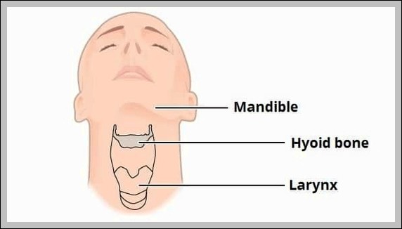
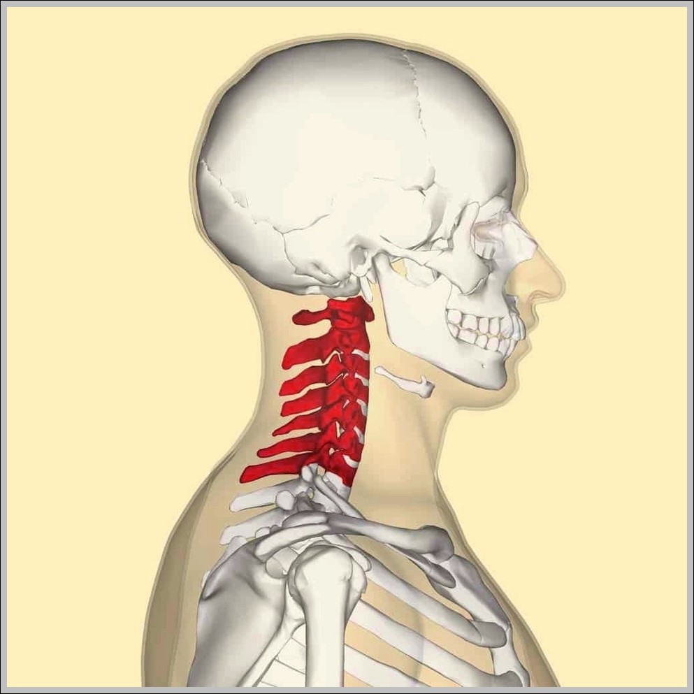
The Position of the Hyoid Bone in the Neck diagram places this small, U-shaped bone right at the C3 level, floating freely without any direct joint connection to other bones.…
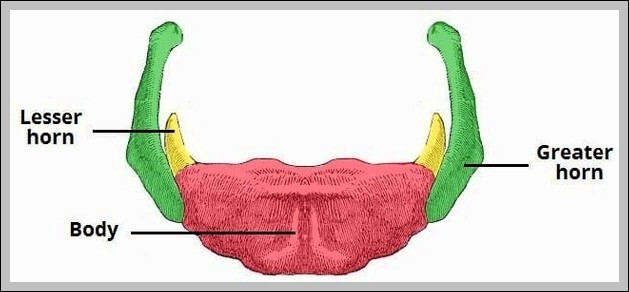
The hyoid bone parts include body (central, C3 level), greater horns (posterior/lateral projections for muscle/ligament attachments), lesser horns (small superior projections for stylohyoid ligament). It floats, anchored by supra/infrahyoid muscles.
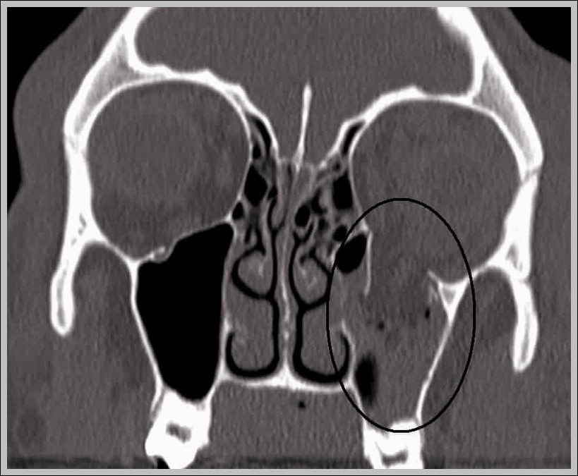
The CT Scan of a Blowout Fracture of the Inferior Wall diagram typically shows orbital floor disruption with herniation of inferior rectus or orbital fat into the maxillary sinus, often…
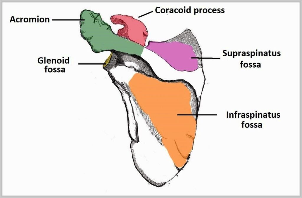
Costal surface of scapula shows subscapular fossa (broad concave area for subscapularis), medial border with superior/inferior angles, costal/vertebral border, and serrated attachments for serratus anterior along medial border.
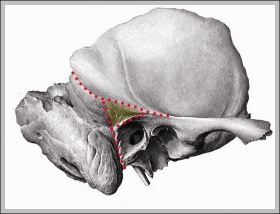
The middle cranial fossa houses the temporal lobes, formed by greater sphenoid wings, squamous temporal, and anterior petrous temporal. Key features include superior orbital fissure, foramen rotundum/ovale/spinosum, carotid canal, and…
The mastoid fossa (antrum) is the largest mastoid air cell, opening into middle ear via aditus ad antrum, lined by mucosa continuous with tympanic cavity. It provides space for middle…
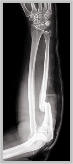
Monteggia fracture: proximal ulna fracture with radial head dislocation (anterior most common). Bado classification types I-IV; requires ORIF for ulna and radial head stability.
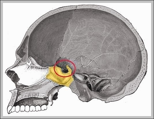
The sella turcica in lateral view is a saddle-shaped depression in sphenoid body housing the pituitary, bounded by tuberculum sellae anteriorly, dorsum sellae posteriorly, and anterior/posterior clinoids.
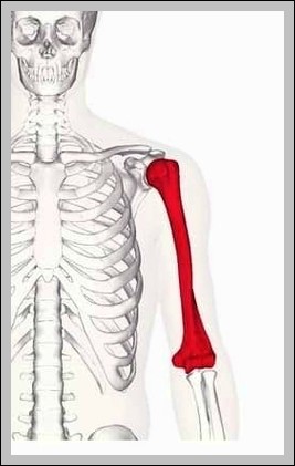
The Anatomical Position of the Humerus diagram places the humerus in standard anatomical position with greater tubercle lateral, capitulum anterior, and trochlea medial. It labels head, surgical/anatomical neck, tubercles, shaft,…
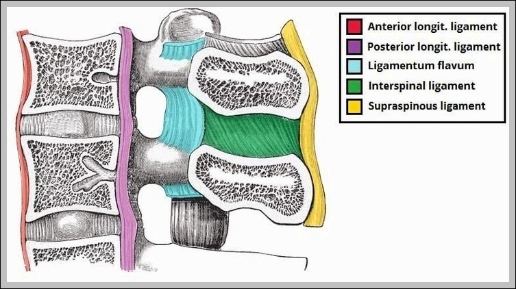
The Ligaments of the Lumbar Spine diagram displays the strong bands that hold the lower vertebrae togetheranterior and posterior longitudinal, ligamentum flavum, interspinous, supraspinous, and others. Seeing them all in…