Skip to content
cropped anatomy systems Anatomy System - Human Body Anatomy diagram and chart images

Human Body Anatomy Diagrams

  • Diagrams
  • Organs
  • Bones
  • Disease
    • Cancer
  • Muscles
  • Cell
  • Health
    • Dental
    • Medical
    • Women
  • Best
  • Home
  • Organs
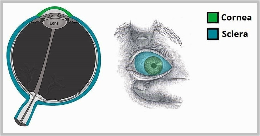
  • The Cornea and Sclera of the Eye Fibrous Layer Diagram
The Cornea and Sclera of the Eye Fibrous Layer Diagram
Posted inOrgans

The Cornea and Sclera of the Eye Fibrous Layer Diagram

Cornea (transparent anterior 1/6) and sclera (opaque posterior 5/6) form fibrous tunic. Cornea avascular, nourished by tears/aqueous; sclera provides shape/protection, continuous at limbus.
Tags:
corneadiagrameyefibrouslayersclera

Post navigation

Previous Post
Parts of the Male Urethra. Diagram Parts of the Male Urethra Diagram
Next Post
Posterior Interosseous Artery Radial Nerve Supintor Diagram Posterior Interosseous Artery Radial Nerve Supintor Diagram
  • Positive Trendlenburg Test Diagram
  • Net Filtration PressureN
  • Anatomical Course of the Subclavian Artery Diagram
  • The Subtalar Joints and Ligament Diagram
  • Articulating Surfaces of the Acromioclavicular Joint Diagram
Copyright 2026 — Anatomy System - Human Body Anatomy diagram and chart images. All rights reserved. Bloghash WordPress Theme
Scroll to Top