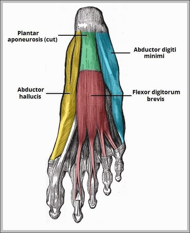
First Layer of Plantar Muscles Diagram

First Layer of Plantar Muscles Diagram

First layer of plantar muscles: abductor hallucis (medial), flexor digitorum brevis (central), abductor digiti minimi (lateral). They arise from calcaneal tuberosity, support medial/longitudinal arch; medial/lateral plantar nerves supply.
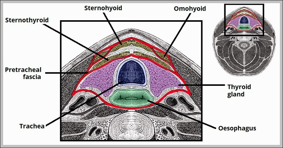
Diagram of the Pretracheal Fascia Layer of the Neck Labelled Diagram

Pretracheal Fascia Layer of the Neck Labelled Diagram

Pretracheal fascia (visceral layer) invests thyroid, trachea, esophagus, infrahyoid muscles; blends with carotid sheath laterally, buccopharyngeal fascia posteriorly. It forms “visceral vascular space” and invests recurrent laryngeal nerves.
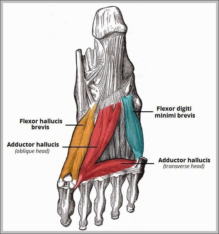
Third Layer of Plantar Muscles Diagram

Third Layer of Plantar Muscles Diagram

Third layer of plantar muscles includes flexor hallucis brevis (to big toe), adductor hallucis (oblique/transverse heads to big toe), and flexor digiti minimi brevis (to little toe). They support toe…