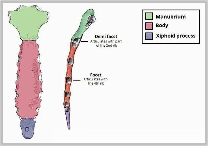
Anatomical Relations of the Prostate Gland Diagram

Anatomical Relations of the Prostate Gland Diagram

Anatomical relations of prostate gland: anterior pubic symphysis/vesicoprostatic space, posterior rectum (separated by Denonvilliers’ fascia), superior bladder neck, inferior urogenital diaphragm, lateral levator ani. Seminal vesicles posterior-superior; urethra passes through.
The Lingual Tonsil Waldeyers Ring Diagram

The Lingual Tonsil Waldeyers Ring Diagram

The lingual tonsil forms part of Waldeyer’s ring on posterior tongue base, composed of lymphoid follicles. It guards oropharynx entrance, enlarges in infection/allergy, visible on endoscopy or lateral neck X-ray.
Tensor Tympani Muscle Middle Ear Diagram

Tensor Tympani Muscle Middle Ear Diagram

Tensor Tympani Muscle Middle Ear diagram shows this tiny muscle originating from the Eustachian tube cartilage and sphenoid, inserting on the malleus handle to dampen loud sounds by tensing the…
Diagram of the Pretracheal Fascia Layer of the Neck Labelled Diagram

Pretracheal Fascia Layer of the Neck Labelled Diagram

Pretracheal fascia (visceral layer) invests thyroid, trachea, esophagus, infrahyoid muscles; blends with carotid sheath laterally, buccopharyngeal fascia posteriorly. It forms “visceral vascular space” and invests recurrent laryngeal nerves.
anatomical terms of location Diagram

anatomical terms of location Diagram

Anatomical terms of location standardize descriptions: superior/inferior, anterior/posterior, medial/lateral, proximal/distal, superficial/deep, palmar/plantar, dorsal/ventral, etc., based on anatomical position.
Elbow3D Diagram

Elbow 3D Diagram

Elbow 3D view shows humeroulnar (hinge), humeroradial (ball/socket), proximal radioulnar (pivot) joints, with trochlea/capitulum, olecranon/coronoid fossae, radial head/annular ligament. It allows flexion/extension and pronation/supination.
Bones of the Middle Cranial Fossa Diagram

Bones of the Middle Cranial Fossa Diagram

The middle cranial fossa houses the temporal lobes, formed by greater sphenoid wings, squamous temporal, and anterior petrous temporal. Key features include superior orbital fissure, foramen rotundum/ovale/spinosum, carotid canal, and…
Structure of the Circle of Willis Diagram

Structure of the Circle of Willis Diagram

The circle of Willis is an anastomotic arterial ring at brain base: anterior cerebral (from internal carotid), anterior communicating, internal carotid, posterior communicating (from posterior cerebral), posterior cerebral (from basilar).…
Diagram of the Descent of the Testis Embryology Diagram

Descent of the Testis Embryology Diagram

Descent of the testis embryologically begins near kidneys at 7-8 weeks, guided by gubernaculum through inguinal canal (transabdominal phase via genitofemoral nerve), then inguinoscrotal phase (hormone-driven, testosterone/insulin-like factor 3) into…
Muscular Triangle of the Neck Diagram

Muscular Triangle of the Neck Diagram

The muscular triangle of the neck is bounded by midline, anterior digastric belly, and superior omohyoid belly, containing infrahyoid muscles (sternohyoid, sternothyroid, thyrohyoid), thyroid gland, trachea, esophagus, and common carotid…
Muscular Attachments to the Mandible Body Ramus Diagram

Muscular Attachments to the Mandible Body Ramus Diagram

Muscular attachments to mandible: body (geniohyoid/genioglossus anterior, mylohyoid lateral, digastric anterior belly), ramus (masseter lateral, medial pterygoid medial, temporalis coronoid). They enable mastication, speech, swallowing.
Anatomical Position and Relations of Pituitary Gland Diagram

Anatomical Position and Relations of Pituitary Gland Diagram

The pituitary gland sits in sella turcica, attached to hypothalamus by infundibulum, surrounded by cavernous sinuses laterally, optic chiasm superiorly, sphenoid sinus inferiorly. Relations affect surgical/transsphenoidal approaches and compression symptoms.
Regional Blood Supply to the Cerebrum Diagram

Regional Blood Supply to the Cerebrum Diagram

Regional blood supply to the cerebrum divides into anterior (anterior cerebral to medial surface), middle (middle cerebral to lateral surface, insula, basal ganglia), and posterior (posterior cerebral to occipital, inferior…