Overview of Parasympathetic Innervation Diagram

Overview of Parasympathetic Innervation Diagram

Parasympathetic innervation originates from cranial nerves (III, VII, IX, X) and sacral S2-S4. Preganglionic fibers synapse in terminal ganglia near targets for “rest and digest” functions: pupil constriction, salivation, lacrimation,…
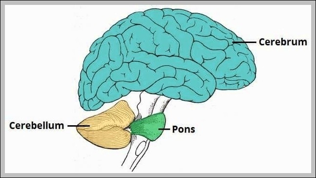
Anatomical Position Cerebellum Diagram

Anatomical Position Cerebellum Diagram

The cerebellum in anatomical position sits in the posterior cranial fossa below the tentorium cerebelli, posterior to the brainstem, with superior surface facing upward toward occipital lobes, inferior surface resting…
Development of the Palate Diagram

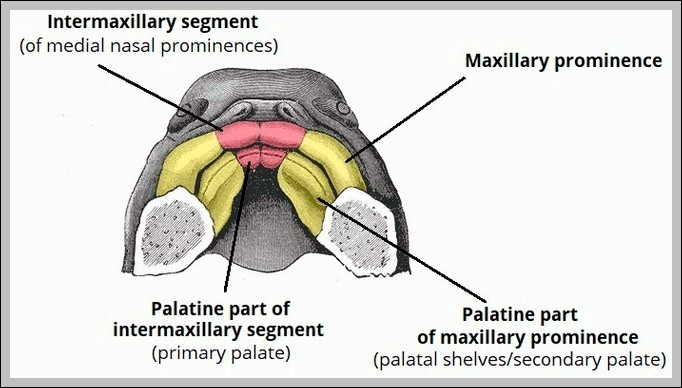
Development of the Palate Diagram

The development of the palate begins with primary palate formation from medial nasal prominences (intermaxillary segment) anteriorly, followed by secondary palate from lateral palatal shelves of maxillary prominences. Shelves grow…
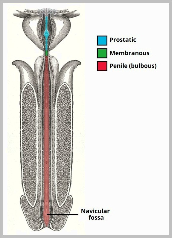
Parts of the Male Urethra. Diagram

Parts of the Male Urethra Diagram

Male urethra parts: prostatic (through prostate), membranous (shortest, through urogenital diaphragm), spongy/penile (longest, through corpus spongiosum to meatus). It conducts urine/semen.
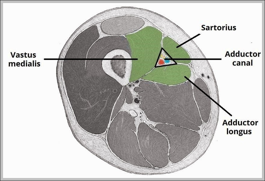
Borders and Contents of the Adductor Canal. Diagram

Borders and Contents of the Adductor Canal Diagram

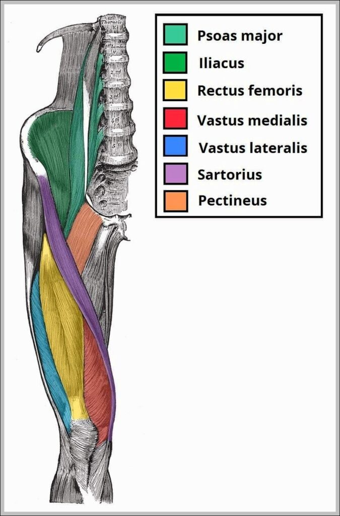
The Borders and Contents of the Adductor Canal diagram defines this subsartorial tunnel from apex of femoral triangle to adductor hiatus: roof (sartorius), anterolateral (vastus medialis), posteromedial (adductors longus/magnus). Contents…
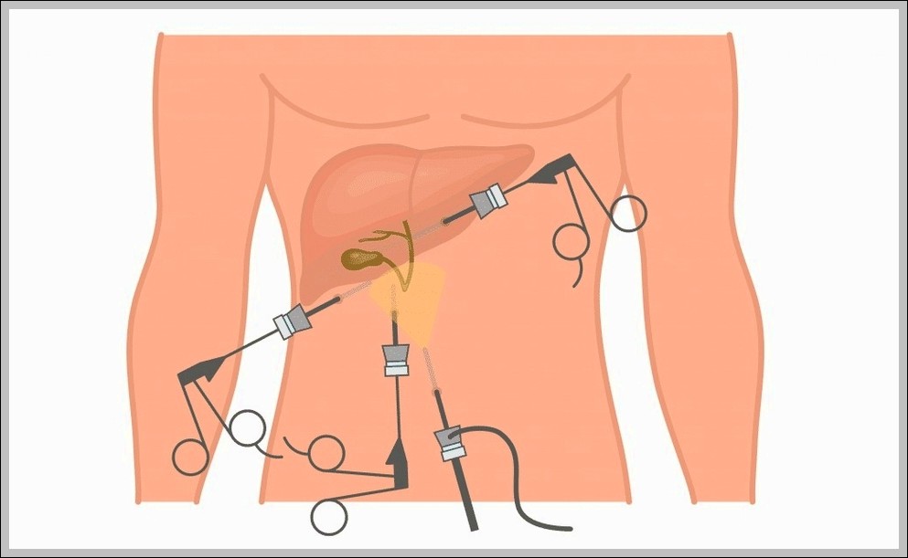
Laparoscopic Cholecystectomy Diagram

Laparoscopic Cholecystectomy Diagram

This Laparoscopic Cholecystectomy diagram shows the key steps of gallbladder removal using small incisions and a camera. It highlights where the trocars go, the tools in action, and important structures…
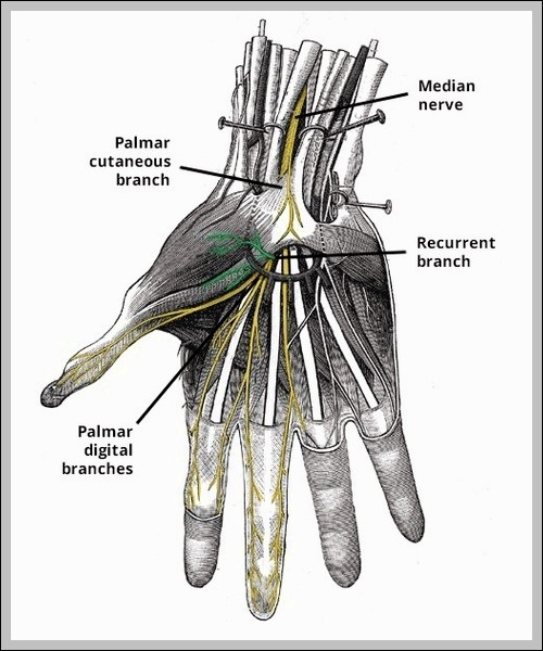
Recurrent Branch of Median Nerve Diagram

Recurrent Branch of Median Nerve Diagram

Recurrent branch of median nerve arises distal to carpal tunnel, loops around flexor pollicis brevis to supply thenar muscles (abductor pollicis brevis, opponens pollicis, superficial flexor pollicis brevis head). It’s…
Bony Landmarks of the Pubis Diagram

Bony Landmarks of the Pubis Diagram

Pubis bony landmarks: body (anterior), superior/inferior rami, pubic tubercle (inguinal ligament attachment), pubic crest, pectineal line, symphyseal surface.
Internal Structure of the Breast Diagram

Internal Structure of the Breast Diagram

Breast internal structure includes 15-20 lobes of glandular tissue draining via lactiferous ducts to nipple, embedded in fat/connective tissue. Cooper’s ligaments provide support; Cooper suspensory.