
The Borders of the Cavernous Sinus diagram gives a clean cross-section view of this important dural venous space next to the pituitary. The walls are clearly markedsuperior, inferior, medial, lateralwhile the internal carotid artery and cranial nerves III, IV, V1, V2, and VI run through or along it. Seeing everything laid out helps explain why infections, clots, or aneurysms here can cause such serious symptoms and why pituitary surgery requires extra caution.
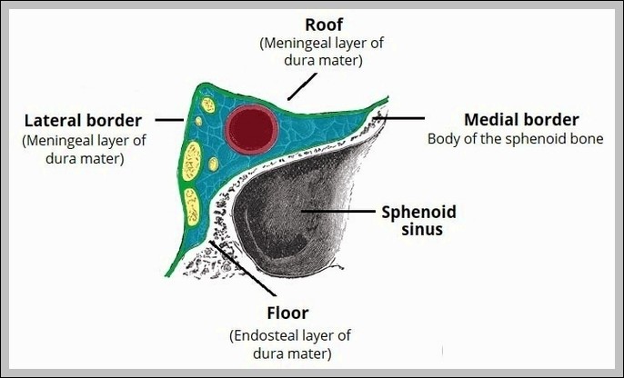
Borders of the Cavernous Sinus Diagram
Posted inMedical
Borders of the Cavernous Sinus Diagram
Post navigation
Previous Post
 Phrenic Nerve Motor Innervation to the Diaphragm Diagram