
Skip to content
cropped anatomy systems Anatomy System - Human Body Anatomy diagram and chart images

Human Body Anatomy Diagrams

  • Diagrams
  • Organs
  • Bones
  • Disease
    • Cancer
  • Muscles
  • Cell
  • Health
    • Dental
    • Medical
    • Women
  • Best
  • Home
  • Organs
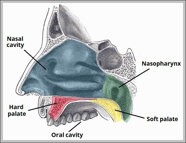
  • The Hard and Soft Palate Diagram
The Hard and Soft Palate Diagram
Posted inOrgans

The Hard and Soft Palate Diagram

Hard palate (maxilla/palatine bones) forms anterior roof of mouth; soft palate (muscular: tensor/levator veli palatini, palatoglossus/pharyngeus) hangs posteriorly with uvula. It separates oral/nasal cavities during swallowing.
Tags:
diagramhardpalatesoft

Post navigation

Previous Post
Anatomical Course of the Inferior Alveolar Nerve and Lingual Nerve Diagram Anatomical Course of the Inferior Alveolar Nerve and Lingual Nerve Diagram
Next Post
Articulations and Parts of the Sternum Diagram Articulations and Parts of the Sternum Diagram
  • Position of the Hyoid Bone in the Neck Diagram
  • Development of the Bladder Ureter Mesonephric Duct Diagram
  • Anatomical Position of the Spleen Diagram
  • Prepubic and Infrapubic Angles of the Male Urethra Diagram
  • Overview of the Anatomical Distribution of the Trigeminal Nerve and its Terminal Branches Diagram
Copyright 2026 — Anatomy System - Human Body Anatomy diagram and chart images. All rights reserved. Bloghash WordPress Theme
Scroll to Top