
Skip to content
cropped anatomy systems

Anatomy System - Human Body Anatomy diagram and chart images

Human Body Anatomy Diagrams

  • Diagrams
  • Organs
  • Bones
  • Disease
    • Cancer
  • Muscles
  • Cell
  • Health
    • Dental
    • Medical
    • Women
  • Best
Top Stories
Microscopic Anatomy of Liver
August 26, 2026
Renin Aldosterone Angiotensin
August 24, 2026
Overview of Parasympathetic Innervation Diagram
August 23, 2026
Anatomical Position Cerebellum Diagram
August 21, 2026
Origin of the Superficial Veins of the Upper Limb Diagram
August 20, 2026
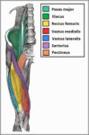
Muscles of the Anterior Thigh Quadriceps Femoris Iliopsoas Sartorius and Pectineus Diagram
August 18, 2026
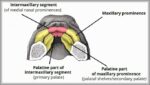
Development of the Palate Diagram
August 17, 2026
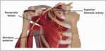
Course of the Superior Thoracic Artery Diagram
August 15, 2026
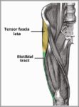
The Tensor Fascia Lata and Iliotibial Tract Diagram
August 14, 2026
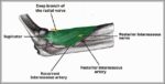
Posterior Interosseous Artery Radial Nerve Supintor Diagram
August 12, 2026
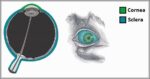
The Cornea and Sclera of the Eye Fibrous Layer Diagram
August 11, 2026
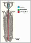
Parts of the Male Urethra. Diagram
Parts of the Male Urethra Diagram
August 9, 2026
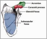
Lateral View of the Scapula Glenoid Fossa Acromion and Coronoid Process Labelled Diagram
August 8, 2026
Location and History of Tonsils
August 6, 2026
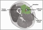
Borders and Contents of the Adductor Canal. Diagram
Borders and Contents of the Adductor Canal Diagram
August 5, 2026
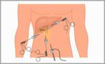
Laparoscopic Cholecystectomy Diagram
August 3, 2026
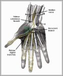
Recurrent Branch of Median Nerve Diagram
August 2, 2026
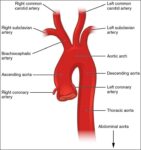
Aorta
Aorta
July 31, 2026
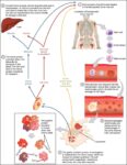
Erythrocyte Life Cycle
July 30, 2026
Spiral Ganglion Neurones Auditory Pathway Diagram
July 28, 2026
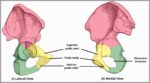
Bony Landmarks of the Pubis Diagram
July 27, 2026
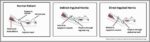
Direct Inguinal Hernia vs Indirect Inguinal Hernia Regional Anatomy Diagram
July 25, 2026
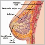
Internal Structure of the Breast Diagram
July 24, 2026
Endocrine System
Endocrine System
July 22, 2026
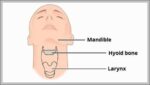
Position of the Hyoid Bone in the Neck Diagram
July 21, 2026
Development of the Bladder Ureter Mesonephric Duct Diagram
July 19, 2026
Anatomical Position of the Spleen Diagram
July 18, 2026
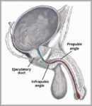
Prepubic and Infrapubic Angles of the Male Urethra Diagram
July 16, 2026
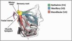
Overview of the Anatomical Distribution of the Trigeminal Nerve and its Terminal Branches Diagram
July 15, 2026
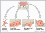
Cellular Components of Bone Diagram
July 13, 2026
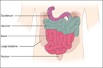
Small IntestineN
Small IntestineN
July 12, 2026
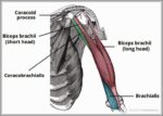
Coracobrachialis Biceps and Brachialis Muscles Diagram
July 10, 2026
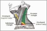
Subdivisions of the Posterior Triangle of the Neck Subclavian Triangle and Occipital Triangle Diagram
July 9, 2026
Neurulation Development of the Central Nervous System Diagram
July 7, 2026
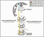
The Spinothalamic Tracts Ascending Sensory 1 Diagram
The Spinothalamic Tracts Ascending Sensory Diagram
July 6, 2026
Course of the Lateral Cutaneous Nerve of the Forearm Diagram
July 4, 2026
Articulations and Parts of the Sternum Diagram
July 3, 2026
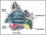
The Hard and Soft Palate Diagram
July 1, 2026
Anatomical Course of the Inferior Alveolar Nerve and Lingual Nerve Diagram
June 30, 2026
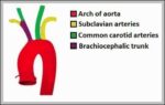
Aorta Diagram
Aorta Diagram
June 28, 2026
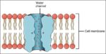
Aquaporin Water Channel
June 27, 2026
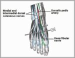
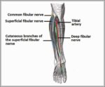
Anatomy of the Nerves of the Dorsum of the Foot Superficial and Deep Fibular Nerves Diagram
June 25, 2026
Anatomical Course of the Common Fibular Nerve and its Terminal Branches Diagram
June 24, 2026
Human Heart Photo
Human Heart Photo
June 22, 2026
Parts of the Hyoid Bone Body Greater Horn Lesser Horn Diagram
June 21, 2026
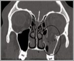
CT Scan of a Blowout Fracture of the Inferior Wall. Diagram
CT Scan of a Blowout Fracture of the Inferior Wall Diagram
June 19, 2026
Heart Valves
Heart Valves
June 18, 2026
Types of Synovial Joint Diagram
June 16, 2026
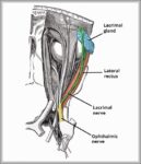
Course of the Lacrimal Nerve Diagram
June 15, 2026
Bony Landmarks of the Costal Surface of the Scapula Diagram
June 13, 2026
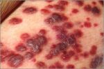
Kaposis Sacroma Lesions
June 12, 2026
Origin of the Blood Supply to the Upper Limb Subclavian Vessels Diagram
June 10, 2026
Medial View of the Tendons of the Muscles in the Post Leg Diagram
June 9, 2026
Anatomical Relations of the Prostate Gland Diagram
June 7, 2026
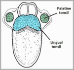
The Lingual Tonsil Waldeyers Ring Diagram
June 6, 2026
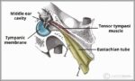
Tensor Tympani Muscle Middle Ear Diagram
June 4, 2026
Granular Leukocytes
Granular Leukocytes
June 3, 2026
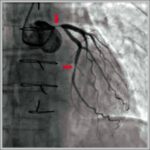
Coronary Angiogram with Narrowing Labelled Diagram
June 1, 2026
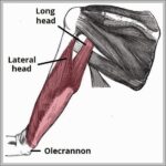
Long and Lateral Heads of the Triceps Brachii Diagram
May 31, 2026
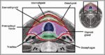
Diagram of the Pretracheal Fascia Layer of the Neck Labelled Diagram
Pretracheal Fascia Layer of the Neck Labelled Diagram
May 29, 2026
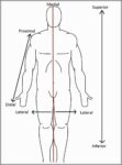
anatomical terms of location Diagram
May 28, 2026
Structure of the Peritoneum and Peritoneal Cavity Diagram
May 26, 2026
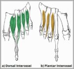
Fourth Layer of the Plantar Muscles Plantar and Dorsal Interossei. Diagram
Fourth Layer of the Plantar Muscles Plantar and Dorsal Interossei Diagram
May 25, 2026
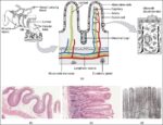
Histology Small IntestinesN
May 23, 2026
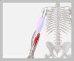
Elbow3D Diagram
Elbow 3D Diagram
May 22, 2026
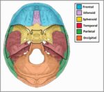
Bones of the Middle Cranial Fossa Diagram
May 20, 2026
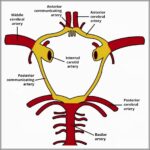
Structure of the Circle of Willis Diagram
May 19, 2026
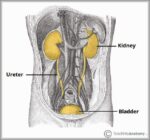
Overview of the Urinary Tract Kidney Ureter and Bladder Diagram
May 17, 2026
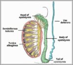
Structure of the Testes and Epididymis Diagram
May 16, 2026
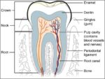
Tooth
Tooth
May 14, 2026
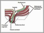
Diagram of the Descent of the Testis Embryology Diagram
Descent of the Testis Embryology Diagram
May 13, 2026
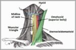
Muscular Triangle of the Neck Diagram
May 11, 2026
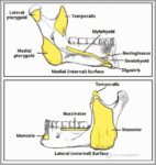
Muscular Attachments to the Mandible Body Ramus Diagram
May 10, 2026
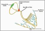
Overview of Conduction Pathway Components Diagram
May 8, 2026
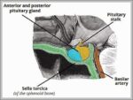
Anatomical Position and Relations of Pituitary Gland Diagram
May 7, 2026
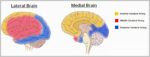
Regional Blood Supply to the Cerebrum Diagram
May 5, 2026
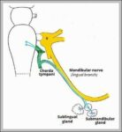
The Submandibular Ganglion and Branches Diagram
May 4, 2026
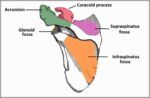
Bony Landmarks of the Posterior Surface of the Scapula Diagram
May 2, 2026
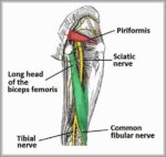
Posterior View of the Lower Limb Anatomical Course of the Sciatic Nerve Diagram
May 1, 2026
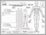
ASIA scale 571 Diagram
ASIA scale 57 Diagram
April 29, 2026
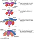
Neurulation
Neurulation
April 28, 2026
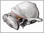
Mastoid Fossa Overview Diagram
April 26, 2026
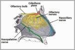
Innervation of the Nasal Septum Olfactory Nasopalatine and Nasociliary Nerves Diagram
April 25, 2026
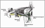
Zygomatic Nerve Course Branches Diagram
April 23, 2026
Radial Nerve Palsy Wristdrop Diagram
April 22, 2026
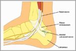
Anatomy of the Medial Calcaneal Nerve Diagram
April 20, 2026
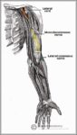
Anatomical Course of the Musculocutaneous Nerve Diagram
April 19, 2026
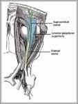
Course of the Supraorbital Nerve Diagram
April 17, 2026
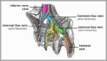
Venous Drainage of the Pelvis Diagram
April 16, 2026
Peristaltic Contractions of the Stomach Diagram
April 14, 2026
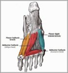
Third Layer of Plantar Muscles Diagram
April 13, 2026
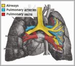
Vasculature of the Lungs Pulmonary Arteries and Veins Diagram
April 11, 2026
Interspinales Muscle of the Back Diagram
April 10, 2026
Posterior Cutaneous Nerve of Thigh Diagram
April 8, 2026
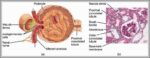
glomerulus Diagram
Glomerulus Diagram
April 7, 2026
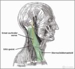
Course of the Great Auricular Nerve Diagram
April 5, 2026
C spine picture Diagram
April 4, 2026
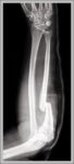
Monteggia Fracture of the Radius and Ulna Diagram
April 2, 2026
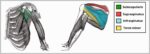
The Rotator Cuff Muscles of the Shoulder Joint Diagram
April 1, 2026
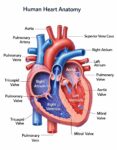
Human Heart Anatomy Diagram
March 31, 2026
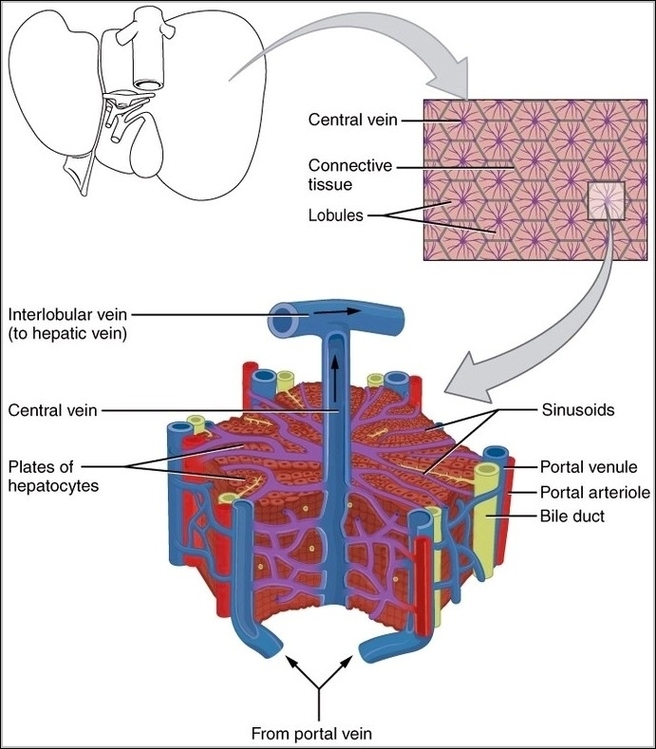
Microscopic Anatomy of Liver
Posted inOrgans

Microscopic Anatomy of Liver

Microscopic anatomy of liver shows hexagonal lobules centered on central vein, with hepatocytes in plates radiating outward, sinusoids lined by fenestrated endothelium and Kupffer macrophages between, portal triads at corners…
Continue Reading
Renin Aldosterone Angiotensin
Posted inMedical

Renin Aldosterone Angiotensin

Renin-angiotensin-aldosterone system activates on low renal perfusion sensed by juxtaglomerular cells releasing renin converting angiotensinogen to angiotensin I, ACE in lungs to II potent vasoconstrictor raising pressure, stimulating aldosterone from…
Continue Reading
Overview of Parasympathetic Innervation Diagram
Posted inMedical

Overview of Parasympathetic Innervation Diagram

Parasympathetic innervation originates from cranial nerves (III, VII, IX, X) and sacral S2-S4. Preganglionic fibers synapse in terminal ganglia near targets for “rest and digest” functions: pupil constriction, salivation, lacrimation,…
Continue Reading
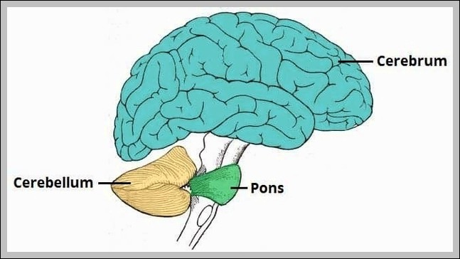
Anatomical Position Cerebellum Diagram
Posted inOrgans

Anatomical Position Cerebellum Diagram

The cerebellum in anatomical position sits in the posterior cranial fossa below the tentorium cerebelli, posterior to the brainstem, with superior surface facing upward toward occipital lobes, inferior surface resting…
Continue Reading
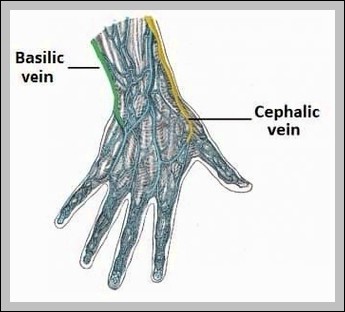
Origin of the Superficial Veins of the Upper Limb Diagram
Posted inMedical

Origin of the Superficial Veins of the Upper Limb Diagram

Superficial upper limb veins originate as dorsal venous arch (cephalic lateral to basilic medial), draining to median cubital at elbow, then axillary/subclavian. They’re key for IV access.
Continue Reading
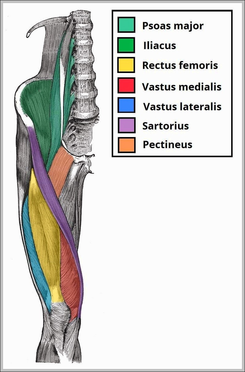
Muscles of the Anterior Thigh Quadriceps Femoris Iliopsoas Sartorius and Pectineus Diagram
Posted inMuscles

Muscles of the Anterior Thigh Quadriceps Femoris Iliopsoas Sartorius and Pectineus Diagram

Anterior thigh muscles: quadriceps femoris (rectus femoris central, vasti around femur), sartorius (crosses from ASIS to pes anserinus), iliopsoas (psoas major/iliacus to lesser trochanter), pectineus (pubis to femur). Femoral nerve…
Continue Reading
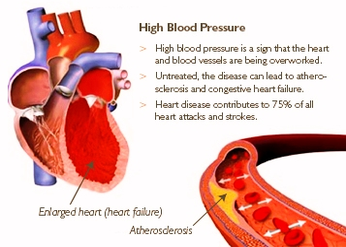
image high blood pressure
Posted inDiagrams Disease

image high blood pressure

image high blood pressure
human skeleton anatomy
Posted inBones Diagrams

human skeleton anatomy

human skeleton anatomy
human leg anatomy diagram
Posted inDiagrams Organs

human leg anatomy diagram

human leg anatomy diagram
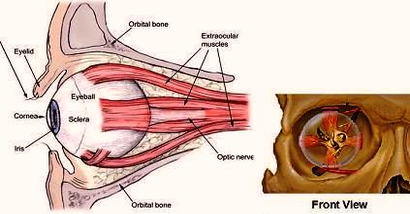
human eye muscles
Posted inMuscles

human eye muscles

human eye muscles
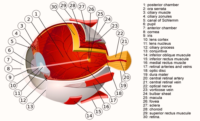
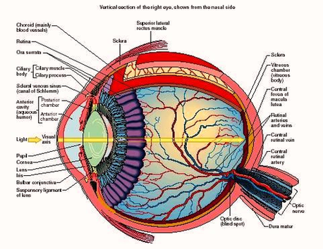
human eye diagram
Posted inOrgans

human eye diagram

human eye diagram
human eye anatomy orbit pic
Posted inDiagrams Organs

human eye anatomy orbit pic

human eye anatomy orbit pic
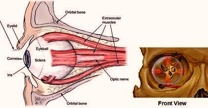
human eye anatomy orbit
Posted inDiagrams Organs

human eye anatomy orbit

human eye anatomy orbit
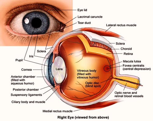
human eye anatomy
Posted inDiagrams Organs

human eye anatomy

human eye anatomy
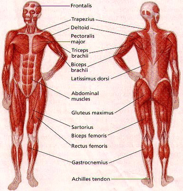
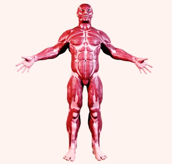
human body muscle diagram
Posted inDiagrams Muscles

human body muscle diagram

human body muscle diagram
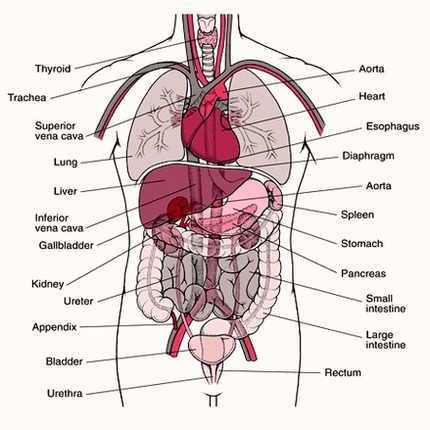
human anatomy organs diagram
Posted inDiagrams Organs

human anatomy organs diagram

human anatomy organs diagram
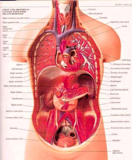
human anatomy organs detailed
Posted inDiagrams Organs

human anatomy organs detailed

human anatomy organs detailed
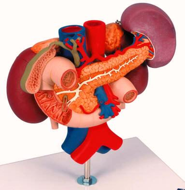
human anatomy organs
Posted inDiagrams Organs

human anatomy organs

human anatomy organs
human anatomy muscular system
Posted inDiagrams Muscles

human anatomy muscular system

human anatomy muscular system
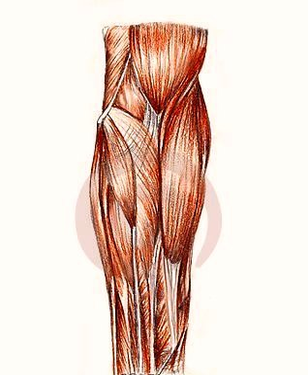
human anatomy muscles arm
Posted inDiagrams Muscles

human anatomy muscles arm

human anatomy muscles arm
human anatomy muscles
Posted inDiagrams Muscles

human anatomy muscles

human anatomy muscles
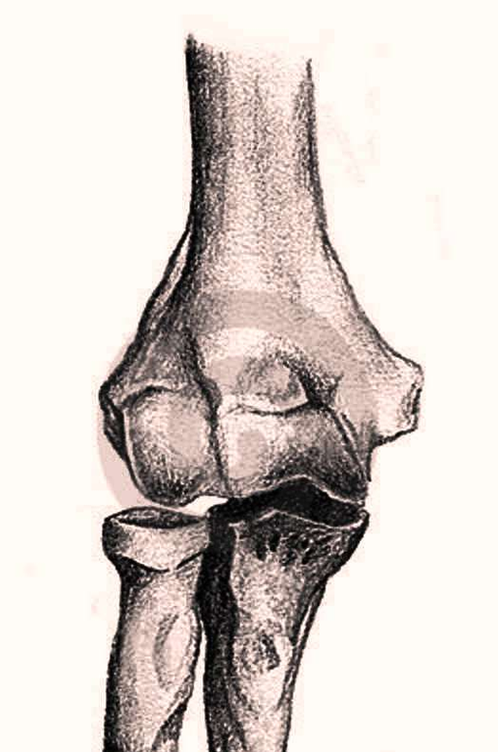
human anatomy bones arm
Posted inBones Diagrams

human anatomy bones arm

human anatomy bones arm
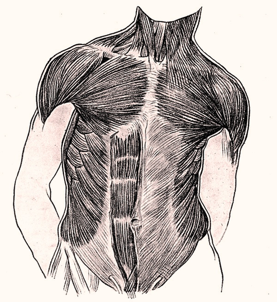
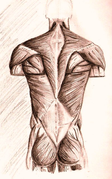
human anatomy back muscles by neilss
Posted inDiagrams Muscles

human anatomy back muscles by neilss

human anatomy back muscles by neilss
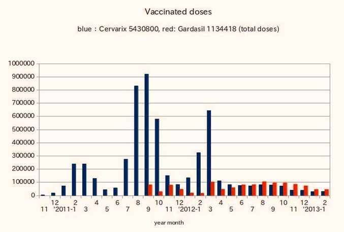
hpv vaccine administered doses figure
Posted inDiagrams Disease Medical

hpv vaccine administered doses figure

hpv vaccine administered doses figure
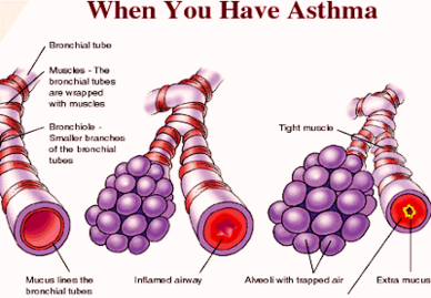
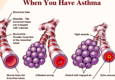
how you get asthma photo
Posted inDiagrams Disease

how you get asthma photo

how you get asthma photo
how you get asthma
Posted inDiagrams Disease

how you get asthma

how you get asthma

Posts pagination

Previous page 1 … 141 142 143 144 145 … 158 Next page
You May Have Missed
Microscopic Anatomy of Liver
Posted inOrgans

Microscopic Anatomy of Liver

Posted by anatomy August 26, 2026
Renin Aldosterone Angiotensin
Posted inMedical

Renin Aldosterone Angiotensin

Posted by anatomy August 24, 2026
Overview of Parasympathetic Innervation Diagram
Posted inMedical

Overview of Parasympathetic Innervation Diagram

Posted by anatomy August 23, 2026
Anatomical Position Cerebellum Diagram
Posted inOrgans

Anatomical Position Cerebellum Diagram

Posted by anatomy August 21, 2026
  • Microscopic Anatomy of Liver
  • Renin Aldosterone Angiotensin
  • Overview of Parasympathetic Innervation Diagram
  • Anatomical Position Cerebellum Diagram
  • Origin of the Superficial Veins of the Upper Limb Diagram
Copyright 2026 — Anatomy System - Human Body Anatomy diagram and chart images. All rights reserved. Bloghash WordPress Theme
Scroll to Top