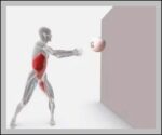
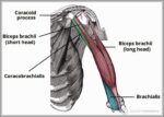
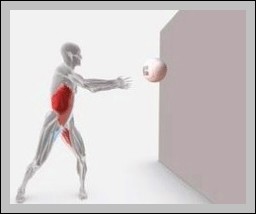
Ball throw mechanics involve shoulder rotation, elbow extension, and wrist snap using rotator cuff, deltoid, triceps, and forearm muscles for power and accuracy.
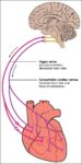
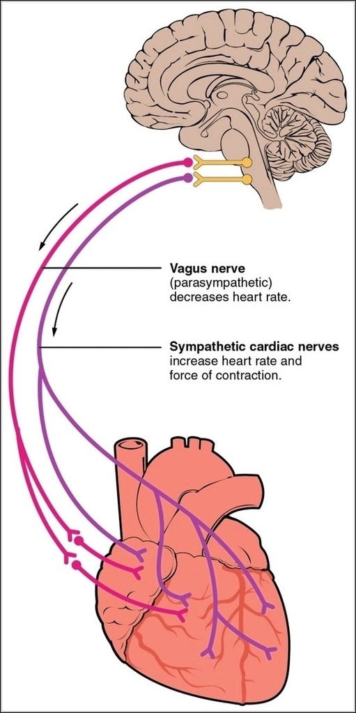
A nervous system diagram of automatic innervation focuses on the autonomic pathways controlling involuntary actions. Sympathetic and parasympathetic branches are labeled, along with target organs like the heart and digestive…
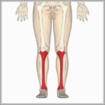
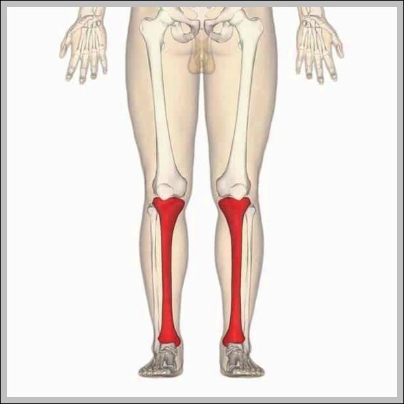
The tibia is the main weight-bearing leg bone with proximal condyles (medial/lateral) for knee joint, tibial tuberosity for patellar ligament, anterior border (shin), interosseous border laterally, and distal medial malleolus.…
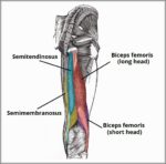
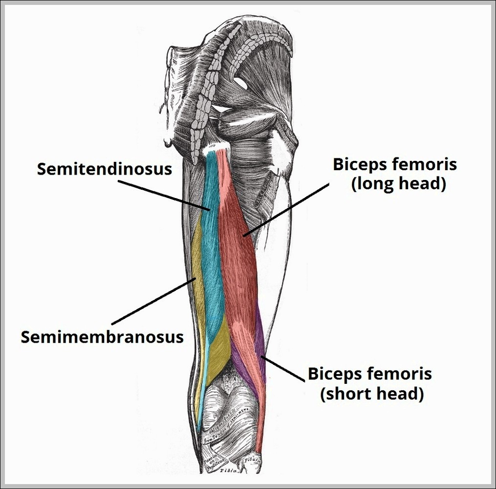
This Muscles of the Posterior Thigh diagram showcases the hamstring groupbiceps femoris (long and short heads), semitendinosus, and semimembranosusoriginating from the ischial tuberosity (except short head from femur) and inserting…
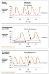
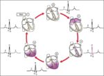
Depolarization in sinus rhythm refers to the orderly spread ??? of electrical impulses through the heart that drives a normal heartbeat, starting from the sinoatrial node, the natural pacemaker located…
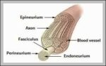
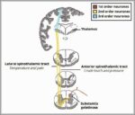
The nervous system or the neural is a complex network of nerve cells or neurons specialized to carry messages. The complexity of the nervous system increases as we move towards…
Last Updated: Jul 16, 2019 The muscular system is responsible for the movement of the human body. Attached to the bones of the skeletal system are about 700 named muscles…
Anatomynote.com found Labelled Diagram Of The Muscles In The Human Body from plenty of anatomical pictures on the internet. We think this is the most useful anatomy picture that you…
Labeled Diagram of the Human Lungs Lungs are an excellent example of how several tissues can be compactly arranged, yet providing a large surface area for gaseous exchange. The current…
Teeth are hard structures found exclusively in all vertebrates. Granted, some invertebrates have analogous “teeth”, but they are quite different in their structure and functions. There are different types of…
The right ventricle is one of the heart’s four chambers. It is located in the lower right portion of the heart below the right atrium and opposite the left ventricle.…
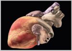
A heart diagram labeled will provide plenty of information about the structure of your heart, including the wall of your heart. The wall of the heart has three different layers,…
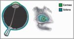
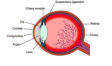
Let’s have a glance at the human eye – it’s structure and functions. A human eye is roughly 2.3 cm in diameter and is almost a spherical ball filled with…
Human eye, specialized sense organ in humans that is capable of receiving visual images, which are relayed to the brain. The anatomy of the eye includes auxiliary structures, such as…
A brief description of the human ear along with a well-labelled diagram is given below for reference. Pinna/auricle is the outermost section of the ear. The external auditory canal links…
(Show more) human ear, organ of hearing and equilibrium that detects and analyzes sound by transduction (or the conversion of sound waves into electrochemical impulses) and maintains the sense of…
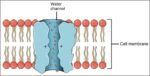
Human Cell Diagram, Parts, Pictures, Structure and Functions. The cell is the basic functional in a human meaning that it is a self-contained and fully operational living entity. Humans are…
These bones are arranged into two major divisions: the axial skeleton and the appendicular skeleton. The axial skeleton runs along the body’s midline axis and is made up of 80…
The human body is made up of several organ systems that work together as one unit. In the pyramid of life that organizes all of the elements of life into…
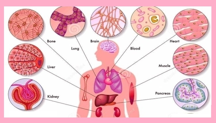
The human body is mainly composed of trillion cells which are considered as the fundamental unit of life. The group of similar cells with similar functions forms a tissue. These…