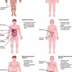
Labeled Diagram of the Human Lungs Lungs are an excellent example of how several tissues can be compactly arranged, yet providing a large surface area for gaseous exchange. The current article provides a labeled diagram of the human lungs as well as a description of the parts and their functions.
The lungs are a pair of spongy, air-filled organs located on either side of the chest (thorax). The trachea (windpipe) conducts inhaled air into the lungs through its tubular branches, called bronchi. The bronchi then divide into smaller and smaller branches (bronchioles), finally becoming microscopic.
The lungs begin at the bottom of your trachea (windpipe). The trachea is a tube that carries the air in and out of your lungs. Each lung has a tube called a bronchus that connects to the trachea.