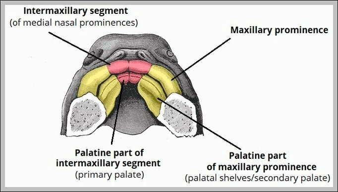
Hard palate (maxilla/palatine bones) forms anterior roof of mouth; soft palate (muscular: tensor/levator veli palatini, palatoglossus/pharyngeus) hangs posteriorly with uvula. It separates oral/nasal cavities during swallowing.
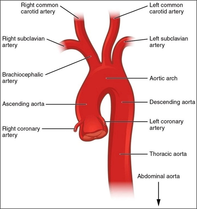
The aorta begins at left ventricle (ascending), arches leftward (arch with three branches), descends thoracic then abdominal to bifurcate at L4 into common iliacs. It supplies all systemic arteries except…
A membrane-level illustration of an aquaporin water channel shows how water crosses cell membranes efficiently. The protein channel is embedded in the lipid bilayer, with water molecules passing single file…
Nerves of dorsum foot include superficial fibular (sensory to dorsum except first webspace), deep fibular (first webspace skin, extensor digitorum brevis), sural (lateral foot). They emerge from anterior/lateral leg compartments.
The common fibular (peroneal) nerve wraps around fibular neck (vulnerable to injury), divides into superficial (lateral compartment, peroneus longus/brevis) and deep (anterior compartment, tibialis anterior, extensors) branches. It supplies dorsiflexion,…
The human heart, about fist-sized and nestled in the mediastinum behind the sternum, has a pointed apex at the left fifth intercostal space and base at the second rib, with…
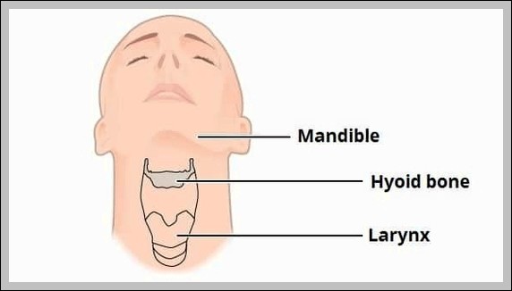
The hyoid bone parts include body (central, C3 level), greater horns (posterior/lateral projections for muscle/ligament attachments), lesser horns (small superior projections for stylohyoid ligament). It floats, anchored by supra/infrahyoid muscles.
The CT Scan of a Blowout Fracture of the Inferior Wall diagram typically shows orbital floor disruption with herniation of inferior rectus or orbital fat into the maxillary sinus, often…
Heart valves ensure one-way blood flow through precisely timed opening and closing. Atrioventricular valvesthe tricuspid on the right and mitral on the lefthang from chordae tendineae anchored to papillary muscles,…
Lacrimal nerve (V1 branch) enters orbit via superior orbital fissure, runs superolateral, supplies lacrimal gland (parasympathetic hitchhike from facial via pterygopalatine ganglion), conjunctiva, upper eyelid skin. It communicates with zygomatic…
Costal surface of scapula shows subscapular fossa (broad concave area for subscapularis), medial border with superior/inferior angles, costal/vertebral border, and serrated attachments for serratus anterior along medial border.
Kaposi's sarcoma lesions appear as purple-red plaques or nodules on skin, mucosa, or viscera, caused by HHV-8 in immunocompromised like AIDS patients, proliferating spindle cells with vascular slits leaking red…
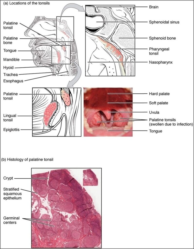
The lingual tonsil forms part of Waldeyers ring on posterior tongue base, composed of lymphoid follicles. It guards oropharynx entrance, enlarges in infection/allergy, visible on endoscopy or lateral neck X-ray.