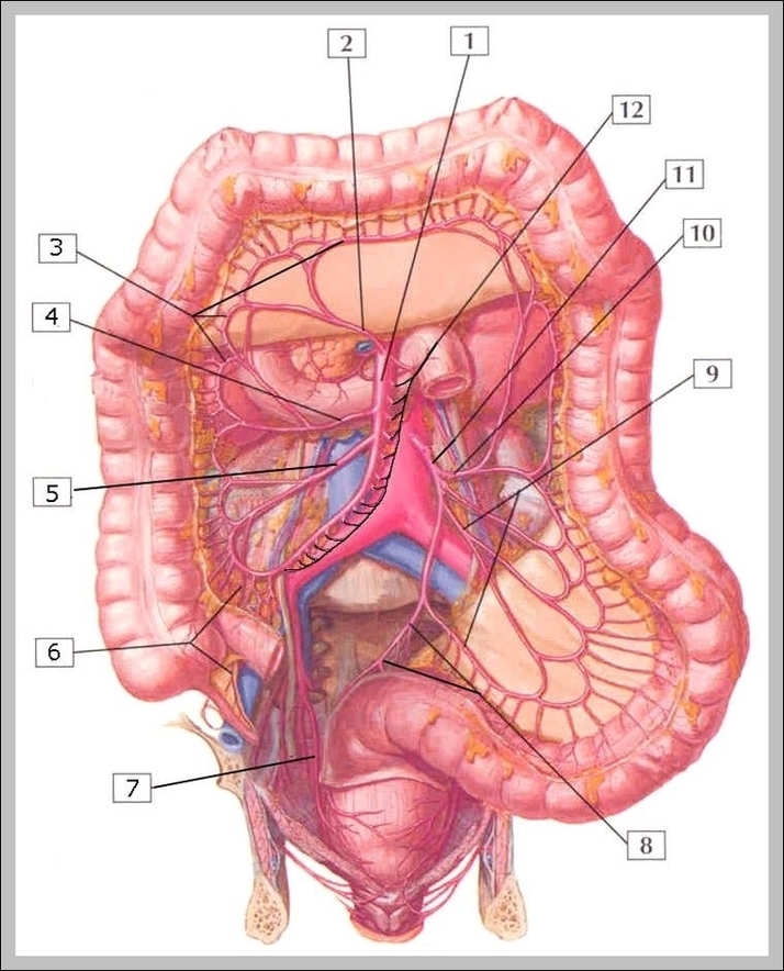
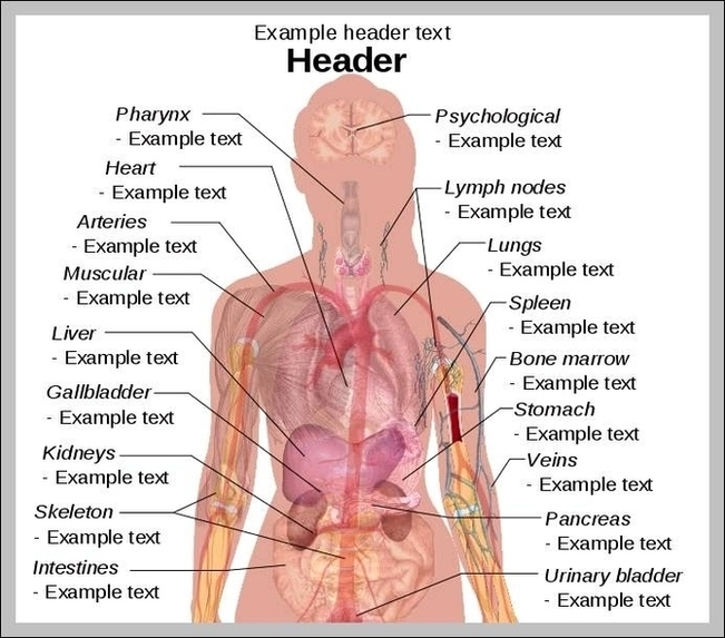
Abdominal Anatomy Picture Image
The main function of the epiglottis is to seal off the windpipe during eating, so that food is not accidentally inhaled. The epiglottis also helps with some aspects of sound production in certain languages.
Epiglottis – dorsal view. The epiglottis is one of the three large unpaired laryngeal cartilages, the other two being the book shaped thyroid and the signet ring shaped cricoid cartilages. The thyroid notch is also termed the laryngeal prominence/Adam’s apple.
Epiglottis – dorsal view. The epiglottis is one of the three large unpaired laryngeal cartilages, the other two being the book shaped thyroid and the signet ring shaped cricoid cartilages. The thyroid notch is also termed the laryngeal prominence/Adam’s apple.