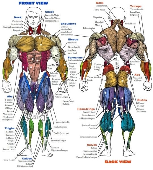
Biceps Brachii Muscle Clarified
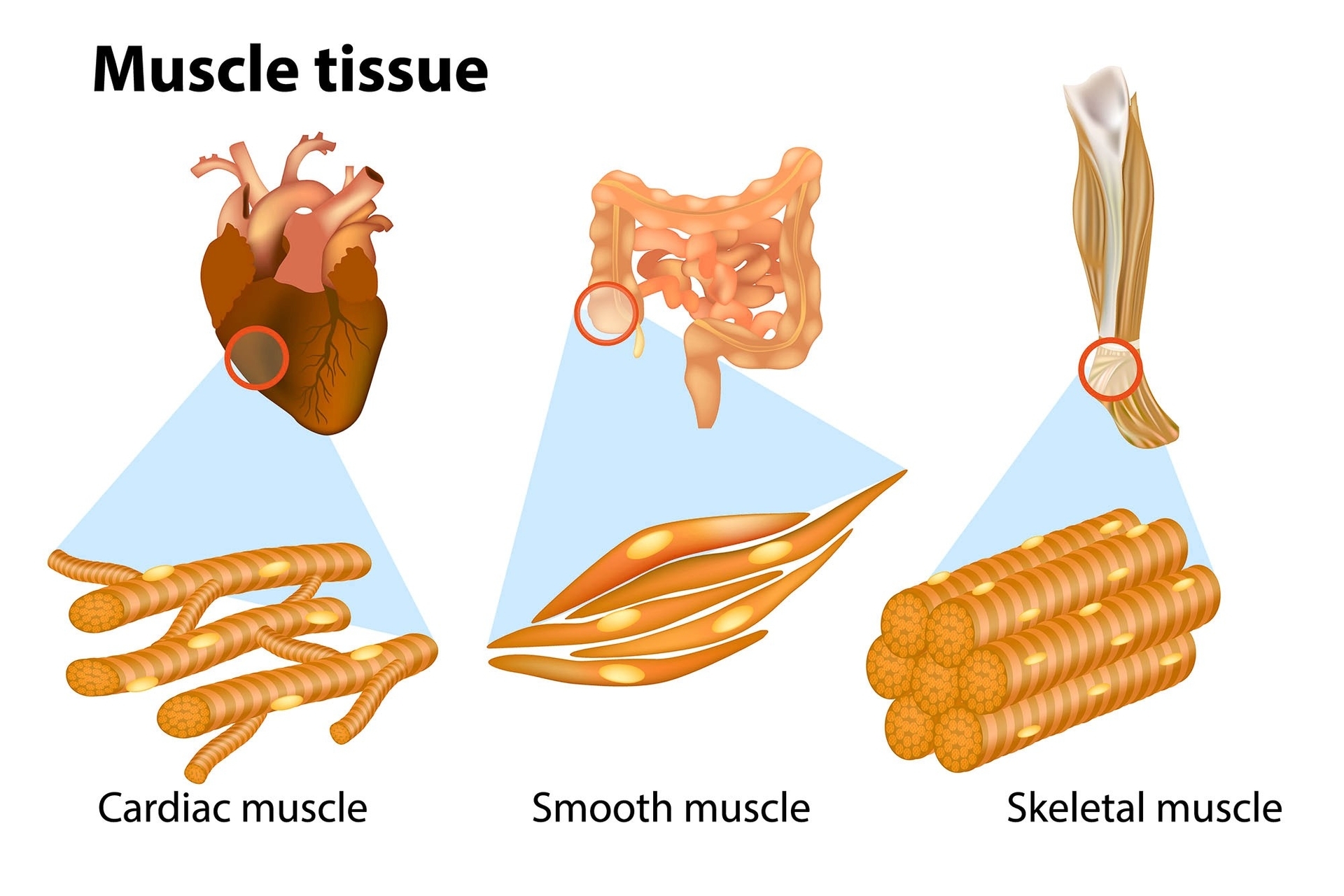
The biceps brachii muscle, commonly known as the biceps, is a prominent skeletal muscle located in the upper arm. Its dual headsthe long head and the short headoriginate from distinct points in the shoulder area, allowing it to participate in both elbow and shoulder movements. Let’s delve into the anatomy and functions of this fascinating muscle.
### Anatomy of the Biceps Brachii Muscle
1. Origins:
– Long Head: The long head originates at the supraglenoid tubercle, situated above the glenoid cavity of the scapula. Remarkably, it lies within the intracapsular space while remaining extrasynovial.
– Short Head: The short head arises at the apex of the coracoid process of the scapula, where it partially blends with the origin tendon of the coracobrachialis. To remember these origins, use the mnemonic: “You walk shorter to a street corner. You ride longer on a superhighway.” The short head originates from the coracoid process, while the long head originates from the supraglenoid cavity. Both heads unite to form a single large muscle belly at the anterior side of the humerus, attaching to the radial tuberosity. Additionally, a fibrous membrane called the bicipital aponeurosis (or lacertus fibrosus) inserts into the deep fascia of the forearm.
2. Insertion:
– The biceps brachii muscle inserts into the radial tuberosity of the radius and the deep fascia of the forearm (specifically at the insertion of the bicipital aponeurosis).
3. Innervation:
– The biceps brachii receives innervation from the musculocutaneous nerve (C5-C6).
4. Blood Supply:
– Branches of the brachial artery supply blood to the muscle.
### Functions of the Biceps Brachii Muscle
1. Flexion of the Forearm at the Elbow Joint:
– The biceps brachii is the prime mover (agonist) responsible for flexing the forearm. When you bend your elbow, the biceps contracts, allowing you to perform actions like lifting objects or curling weights.
2. Supination of the Forearm:
– Supination involves rotating the forearm so that the palm faces upward. The biceps assists in this movement by working in conjunction with other muscles.
3. Weak Flexion of the Arm at the Glenohumeral Joint:
– Although its primary role is elbow flexion, the biceps also contributes to weak flexion of the arm at the shoulder joint (glenohumeral joint).
### Clinical Notes
– Biceps Tendon Injuries: Tears or inflammation of the biceps tendon can occur due to trauma or overuse. These injuries may lead to pain, weakness, and limited range of motion.
– Biceps Brachii in Strength Training: Bodybuilders and fitness enthusiasts often target the biceps brachii during exercises like bicep curls to enhance arm aesthetics and strength.
In summary, the biceps brachii muscle, with its dual heads and unique structure, plays a crucial role in everyday movements and athletic endeavors. Whether you’re lifting groceries or hitting the gym, this muscle quietly supports your actions, making it a true unsung hero of the upper arm..
Biceps Brachii Muscle Clarified Diagram - Biceps Brachii Muscle Clarified Chart - Human anatomy diagrams and charts explained. This anatomy system diagram depicts Biceps Brachii Muscle Clarified with parts and labels. Best diagram to help learn about health, human body and medicine.