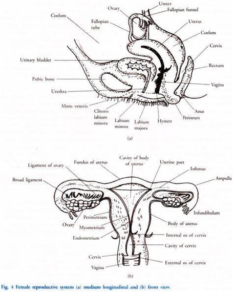
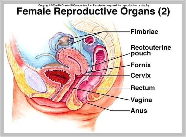
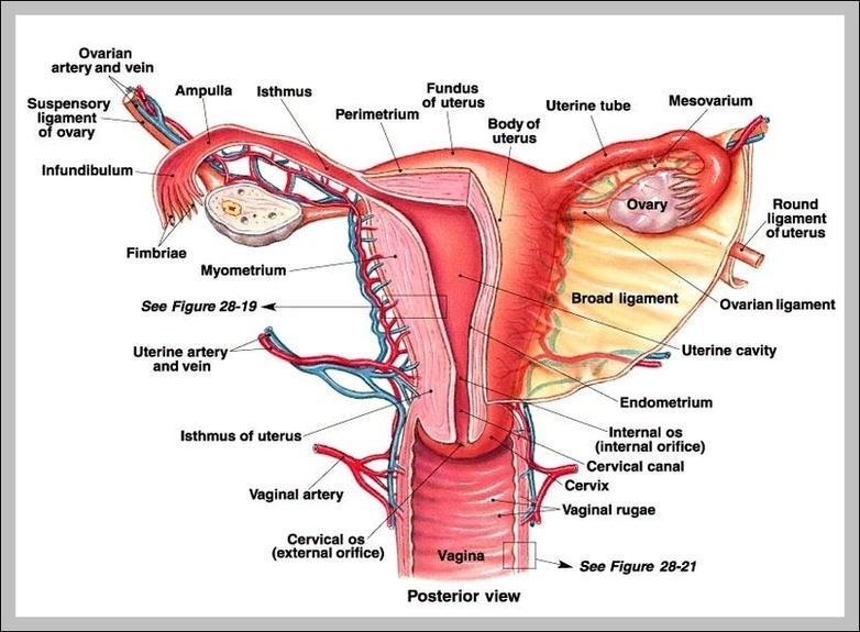
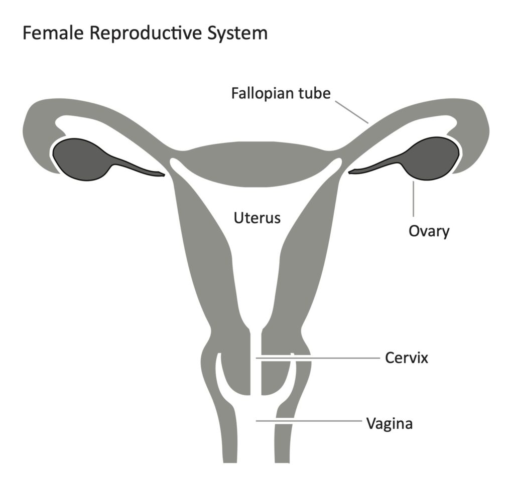
Human Anatomy For Muscle, Reproductive, And Skeleton Represented

Human Anatomy: Muscle, Reproductive, and Skeletal Systems

Muscular System

The muscular system is responsible for movement, posture, and balance. It consists of three types of muscle tissue: skeletal, smooth, and cardiac. Skeletal muscles, numbering over 600, create movement in the body. They work in groups to move the skeleton, with movements being nearly involuntary, yet requiring conscious effort. Cardiac muscle, found in the heart, is responsible for the rhythmic contractions that pump blood throughout the body. Smooth muscle, found in the walls of hollow organs and blood vessels, propels substances through the body.

Reproductive System

The human reproductive system allows for the production and fertilization of gametes, leading to the creation of offspring[^10^]. In males, the testes produce sperm, and the penis delivers the sperm for potential fertilization[^10^]. In females, the ovaries produce eggs, the uterus houses the developing fetus, and the breasts produce milk for the newborn. The reproductive process involves the release of an egg, internal fertilization by sperm, transport of the fertilized egg to the uterus, implantation in the uterine wall, gestation, birth, and postnatal care[^10^].
keletal System

The skeletal system serves as the body’s framework, providing support, protection, and enabling motion?. It consists of 206 bones, divided into the axial and appendicular skeletons?. The axial skeleton includes the vertebral column and much of the skull, providing support and protection for the body’s central parts?. The appendicular skeleton includes the pelvic and pectoral girdles and the bones of the limbs?. The skeletal system also includes ligaments, which attach bone to bone, and cartilage, which provides padding between bones?.

In conclusion, the muscular, reproductive, and skeletal systems each play crucial roles in the human body. The muscular system enables movement and maintains posture, the reproductive system allows for the continuation of human species, and the skeletal system provides structural support and protection for the body’s organs. Each system, with its unique structure and function, contributes to the overall health and well-being of an individual..

Human Anatomy For Muscle, Reproductive, And Skeleton Represented Diagram - Human Anatomy For Muscle, Reproductive, And Skeleton Represented Chart - Human anatomy diagrams and charts explained. This anatomy system diagram depicts Human Anatomy For Muscle, Reproductive, And Skeleton Represented with parts and labels. Best diagram to help learn about health, human body and medicine.

Human Anatomy For Muscle, Reproductive, And Skeleton Represented

Human Anatomy For Muscle, Reproductive, And Skeleton

Human Anatomy: Muscle, Reproductive, and Skeletal Systems

Muscular System
The muscular system is responsible for movement, posture, and balance. It consists of three types of muscles: skeletal, smooth, and cardiac.

– Skeletal Muscles: These muscles are attached to the bones by tendons and work in groups to move the skeleton. They make up about 40% of a person’s body weight.
– Smooth Muscles: Found in the walls of hollow organs, blood vessels, and respiratory passageways, they contract in response to stimuli and nerve impulses.
– Cardiac Muscles: These muscles make up the walls of the heart and are responsible for the rhythmic contractions that pump blood through the body.

Reproductive System
The reproductive system is responsible for human reproduction and bearing live offspring?. It includes both internal and external genitalia?.

– Male Reproductive System: The male reproductive system includes the testes that produce sperm and a penis for delivery?. The sperm mature in the testes and then enter the epididymis for further maturation?.
– Female Reproductive System: The female reproductive system includes the ovaries that produce eggs, a uterus for baby development, and breasts for milk production?. The ovum is released at a specific time in the reproductive cycle for internal fertilization by sperm cells?.
keletal System
The skeletal system serves as a framework for the body, providing shape, stability, and protection of internal organs. It consists of 206 bones, ligaments, and cartilages.

– Axial Skeleton: This includes the vertebral column (the spine) and much of the skull, providing the main support of the trunk.
– Appendicular Skeleton: This includes the pelvic (hip) and pectoral (shoulder) girdles and the bones and cartilages of the limbs.

In conclusion, the muscular, reproductive, and skeletal systems play crucial roles in the functioning of the human body. Each system has a unique structure and specific role, contributing to our movement, reproduction, and structural support.

Human Anatomy For Muscle, Reproductive, And Skeleton Diagram - Human Anatomy For Muscle, Reproductive, And Skeleton Chart - Human anatomy diagrams and charts explained. This anatomy system diagram depicts Human Anatomy For Muscle, Reproductive, And Skeleton with parts and labels. Best diagram to help learn about health, human body and medicine.

Human Anatomy For Muscle, Reproductive, And Skeleton

Female Reproductive System Anatomy Diagram Image

Female reproductive system. Genital organs. Human anatomy. Cervix, ovaries, fallopian tubes. Vector illustration. female reproductive system diagram stock illustrations Uterus. Female reproductive system. Genital organs. Human anatomy. Set of Female reproductive system uterus in different styles and cross sections. Front view in a cut.
465 female anatomy diagram stock photos and images available, or start a new search to explore more stock photos and images. Figures illustrating the difference between a natural waist and a constricted waist deformed through the use of corsets, from a published essay by…

Female Reproductive System Anatomy Diagram Image Diagram - Female Reproductive System Anatomy Diagram Image Chart - Human anatomy diagrams and charts explained. This anatomy system diagram depicts Female Reproductive System Anatomy Diagram Image with parts and labels. Best diagram to help learn about health, human body and medicine.

Female Reproductive System Anatomy Diagram Image

What Is The Female Reproductive System Image

Female Reproductive System 1 The female reproductive anatomy includes both external… 2 Females of reproductive age… 3 Follicular phase. This phase starts on the first day of your period. 4 Ovulatory phase. The ovulatory phase (ovulation)…
3D computer graphic sagittal section of female reproductive system with labels. The female reproductive system (or female genital system) contains two main parts: the uterus, which hosts the developing fetus, produces vaginal and uterine secretions Anterior view of uterus, fallopian tube, ovary, cervix.

What Is The Female Reproductive System Image Diagram - What Is The Female Reproductive System Image Chart - Human anatomy diagrams and charts explained. This anatomy system diagram depicts What Is The Female Reproductive System Image with parts and labels. Best diagram to help learn about health, human body and medicine.

What Is The Female Reproductive System Image

Anatomy Of Reproductive System Image

Anatomy Of Reproductive System Image Diagram - Anatomy Of Reproductive System Image Chart - Human anatomy diagrams and charts explained. This anatomy system diagram depicts Anatomy Of Reproductive System Image with parts and labels. Best diagram to help learn about health, human body and medicine.

Anatomy Of Reproductive System Image

Picture Of Reproductive System Image

Picture Of Reproductive System Image Diagram - Picture Of Reproductive System Image Chart - Human anatomy diagrams and charts explained. This anatomy system diagram depicts Picture Of Reproductive System Image with parts and labels. Best diagram to help learn about health, human body and medicine.

Picture Of Reproductive System Image

Reproductive System Function Image

What Is the Function of the Reproductive System? In a large number of species, the reproductive system hosts organs and systems, such as gonads, needed for successful reproduction and also secretes sex hormones such as progesterone and testosterone, notes Wikipedia.
3D computer graphic sagittal section of female reproductive system with labels. The female reproductive system (or female genital system) contains two main parts: the uterus, which hosts the developing fetus, produces vaginal and uterine secretions Anterior view of uterus, fallopian tube, ovary, cervix.

Reproductive System Function Image Diagram - Reproductive System Function Image Chart - Human anatomy diagrams and charts explained. This anatomy system diagram depicts Reproductive System Function Image with parts and labels. Best diagram to help learn about health, human body and medicine.

Reproductive System Function Image

Males Reproductive System Image

3D computer graphic cross section of male reproductive system with labels. Male reproductive system The human male reproductive system consists of a number of sex organs that form a part of the human reproductive process.
Browse 12,801 male reproductive organ stock photos and images available, or search for scrotum or testis to find more great stock photos and pictures. Illustration of the anatomy of the male reproductive system on a white background
You have several internal organs — also called accessory organs — that play a big part in the male reproductive system. These organs include: Vas deferens: The vas deferens is a long, muscular tube that travels from the epididymis into the pelvic cavity, to just behind the bladder.

Males Reproductive System Image Diagram - Males Reproductive System Image Chart - Human anatomy diagrams and charts explained. This anatomy system diagram depicts Males Reproductive System Image with parts and labels. Best diagram to help learn about health, human body and medicine.

Males Reproductive System Image

Reproductive Male Diagram

The male reproductive system consists of a pair of the testis, the duct system, accessory glands, and external genitalia. Let us understand in detail the male reproductive system through the diagram given below: The testicles are oval and located outside the abdominal cavity within a scrotum pouch.
Unlike Female Reproductive System, the male reproductive system is an external body part. Apart from these components, the male reproductive system also includes seminal vesicles, prostate glands, and bulbourethral glands, which perform various functions in the body.
Sexual reproduction is a biparental type of reproduction that involves the production of gametes and there is a fusion of male and female gametes to produce a diploid zygote. What is Male Reproductive System? The human male reproductive system is a group of organs that takes part in the process of reproduction and produces gametes.

Reproductive Male Diagram Diagram - Reproductive Male Diagram Chart - Human anatomy diagrams and charts explained. This anatomy system diagram depicts Reproductive Male Diagram with parts and labels. Best diagram to help learn about health, human body and medicine.

reproductive male diagram

Reproductive Female Diagram

The female reproductive system is composed of a pair of ovaries along with oviducts, vagina, cervix, uterus, and the external genitalia that are located in the pelvic region.
Female anatomy refers to the internal and external structures of the reproductive and urinary systems. Reproductive anatomy aids with sexual pleasure, getting pregnant, and breastfeeding a baby. The urinary system helps rid the body of toxins through urination (peeing).
What do the reproductive organs look like (picture)? The uterus, or womb, is a hollow organ located centrally in the pelvis. It houses the developing fetus during pregnancy. The lower portion of the uterus is called the cervix and opens into the vagina, or birth canal.

Reproductive Female Diagram Diagram - Reproductive Female Diagram Chart - Human anatomy diagrams and charts explained. This anatomy system diagram depicts Reproductive Female Diagram with parts and labels. Best diagram to help learn about health, human body and medicine.

reproductive female diagram