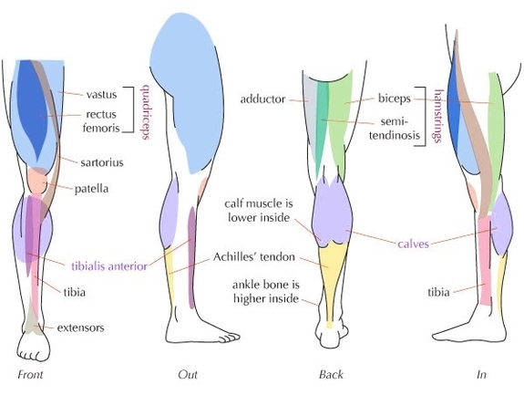
The human leg, a complex structure with numerous muscles, plays a pivotal role in body movement and support. The majority of leg muscles are considered long muscles, stretching great distances to move skeletal bones and facilitate body movement.
Upper Leg Muscles
The upper leg comprises the quadriceps and hamstrings. The quadriceps, the body’s strongest and leanest muscles, are major extensors of the knee. They include:
1. Vastus lateralis: The largest of the quadriceps, it extends from the top of the femur to the kneecap.
2. Vastus medialis: A teardrop-shaped muscle of the inner thigh, it attaches along the femur and down to the inner border of the kneecap.
3. Vastus intermedius: Located between the vastus medialis and the vastus lateralis at the front of the femur, it is the deepest of the quadriceps muscles.
4. Rectus femoris: This muscle attaches to the kneecap.
The hamstrings, three muscles at the back of the thigh, affect hip and knee movement. They include:
1. Biceps femoris: This long muscle flexes the knee.
2. Semimembranosus: This long muscle extends from the pelvis to the tibia.
3. Semitendinosus: This muscle extends the thigh and flexes the knee.
Lower Leg Muscles
The lower leg muscles, supported by the fibula and the tibia (shinbone), are pivotal to movement of the ankle, foot, and toes. Some of the major muscles of the calf include:
1. Gastrocnemius (calf muscle): One of the large muscles of the leg, it connects to the heel. It flexes and extends the foot, ankle, and knee.
2. Soleus: This muscle extends from the back of the knee to the heel. It is important in walking and standing.
3. Plantaris: This small, thin muscle is absent in about 10 percent of people. The gastrocnemius muscle supersedes its function.
The Achilles tendon, connecting the plantaris, gastrocnemius, and soleus muscles to the heel bone, stores the elastic energy needed for running, jumping, and other physical activity.
Functional Groups
The leg muscles are organized into three groups: anterior (dorsiflexor) group, posterior (plantar flexor) group, and lateral (fibular) group. These groups produce different movements in the ankle and foot, crucial for activities such as walking, running, and dancing.
The anterior group, including the tibialis anterior, extensor digitorum longus, fibularis tertius, and extensor hallucis longus, primarily produces dorsiflexion of the foot at the ankle joint. The posterior group, comprising the gastrocnemius, plantaris, soleus