Posted inMedical
Anatomy of the Nerves of the Dorsum of the Foot Superficial and Deep Fibular Nerves Diagram
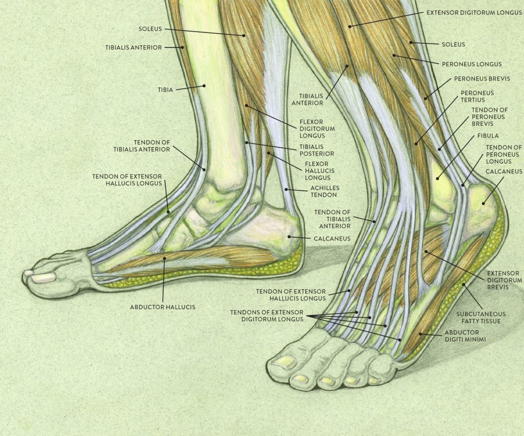
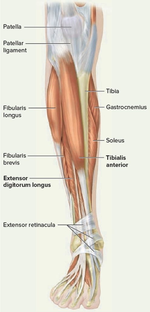
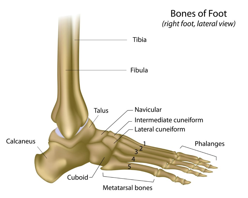
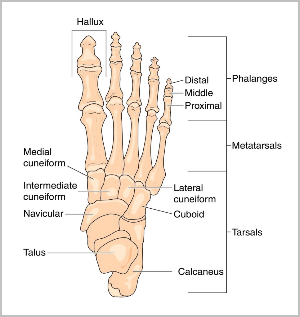
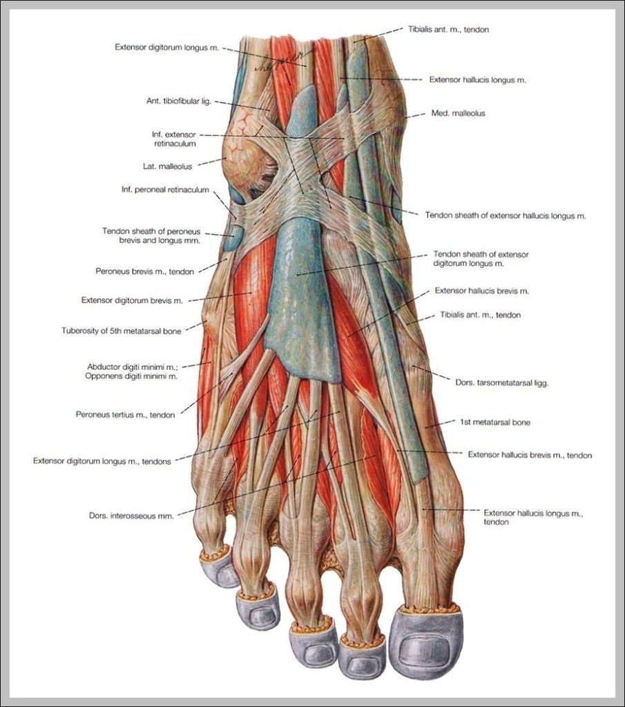
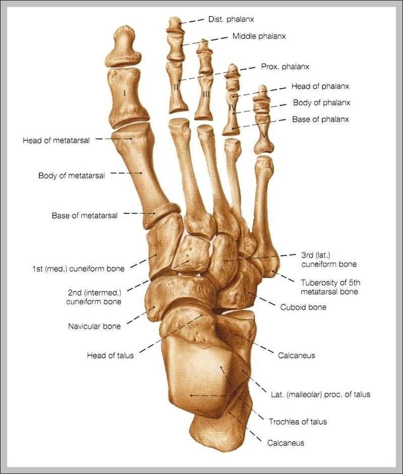
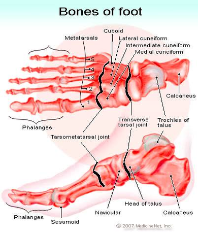
Nerves of dorsum foot include superficial fibular (sensory to dorsum except first webspace), deep fibular (first webspace skin, extensor digitorum brevis), sural (lateral foot). They emerge from anterior/lateral leg compartments.