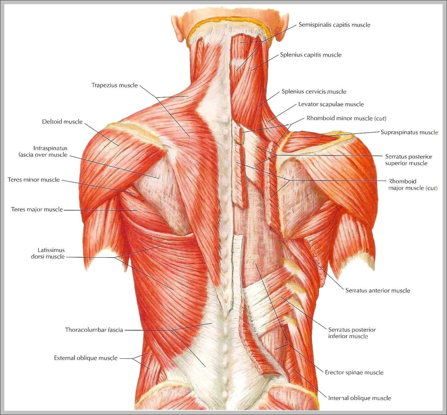
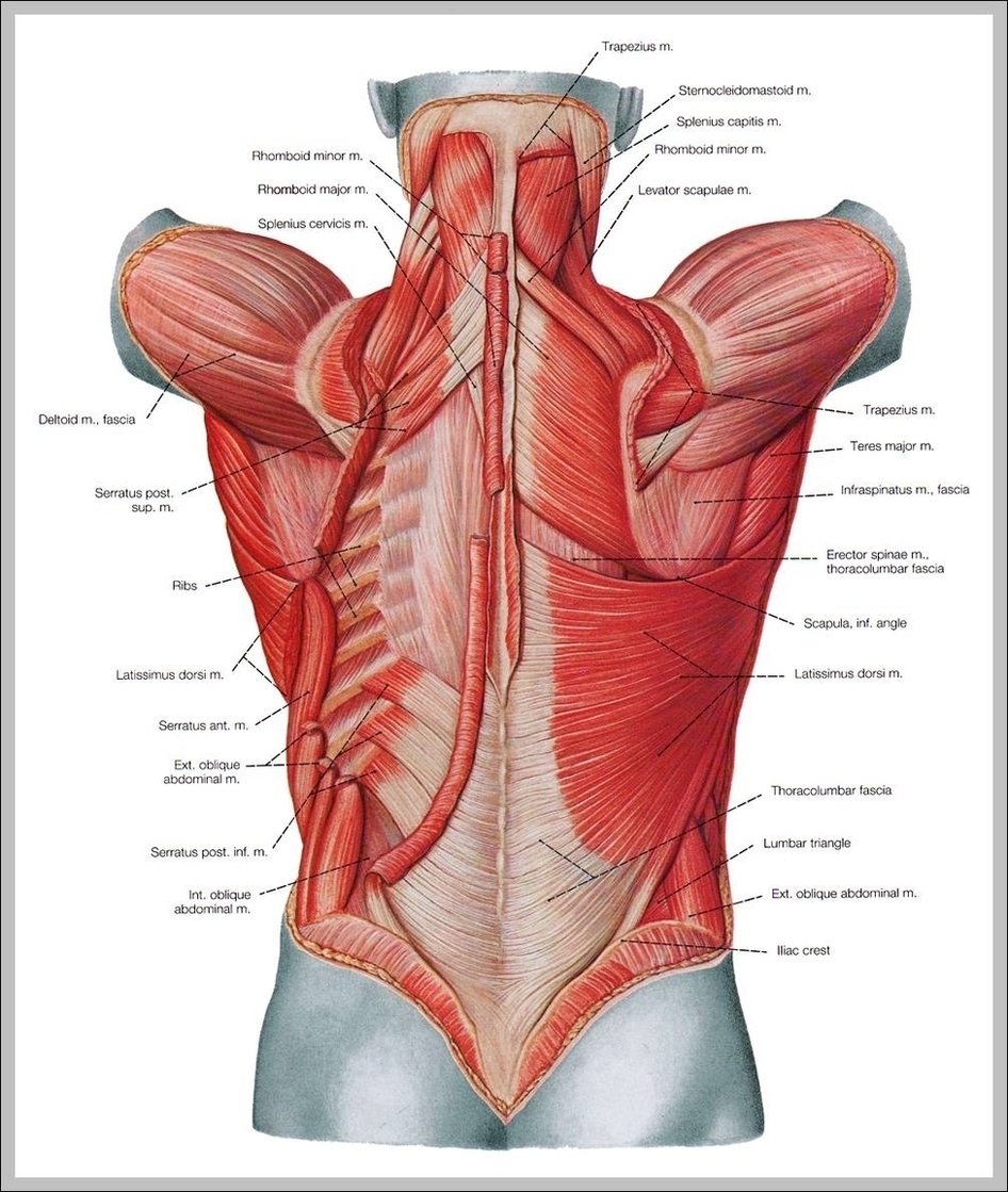
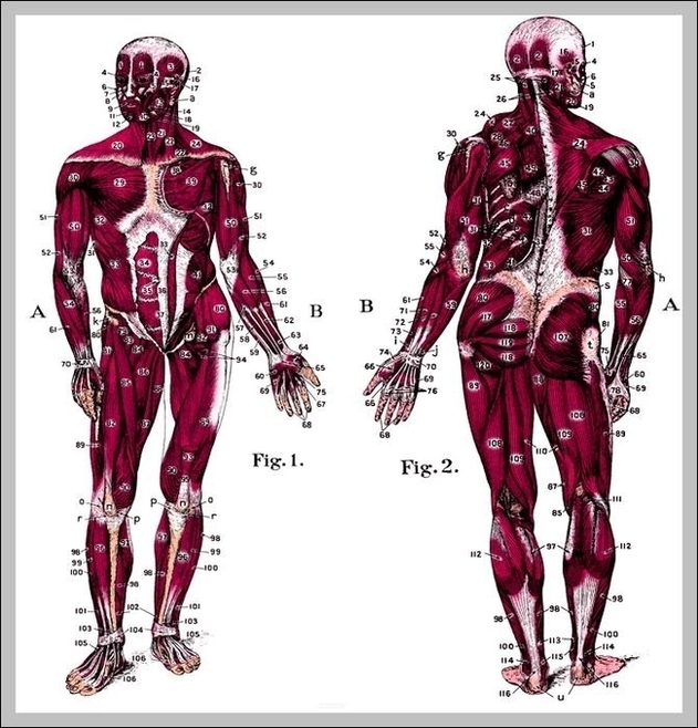
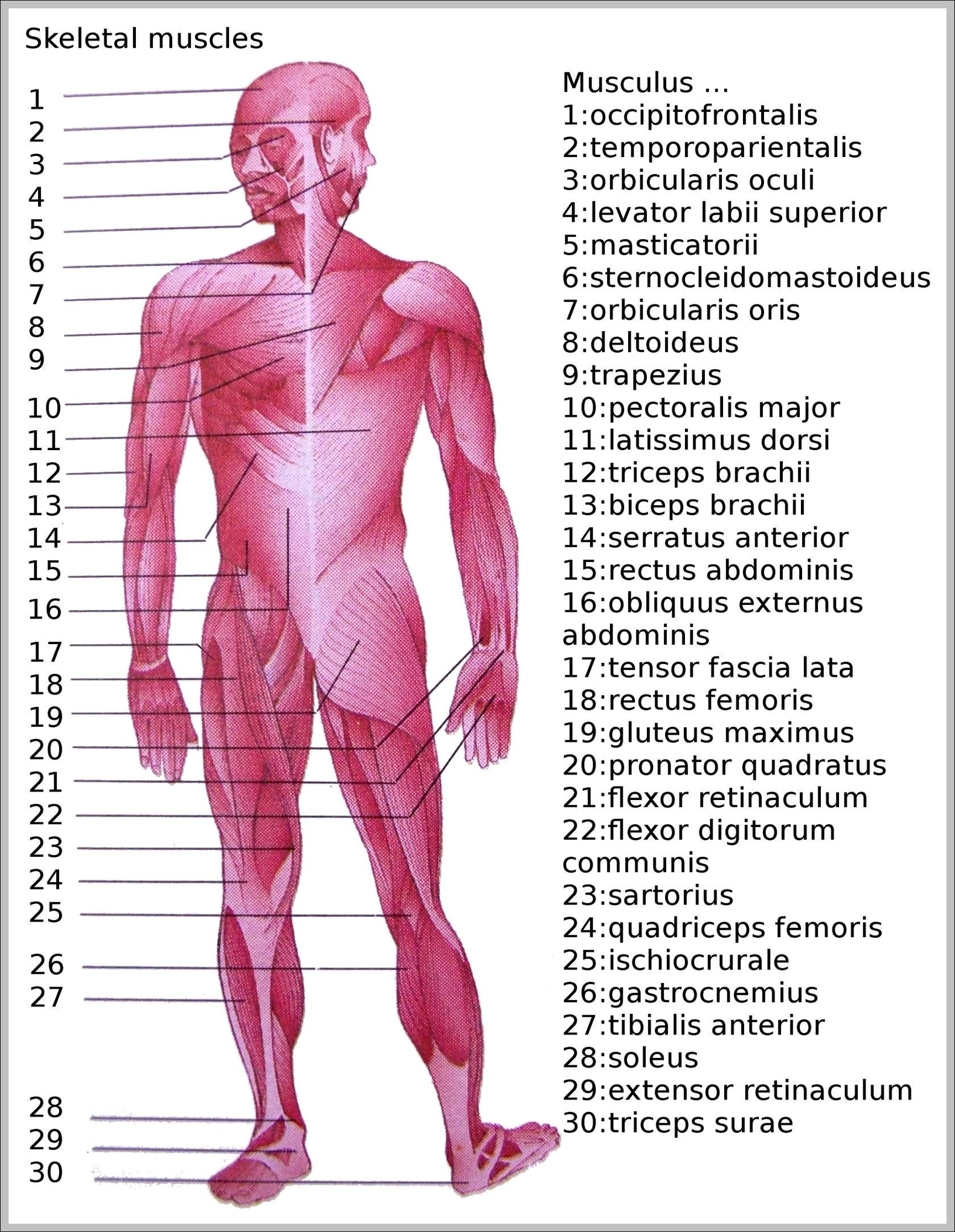
Upper Back Muscle Anatomy Image
Your back consists of a complex array of bones, discs, nerves, joints, and muscles. The muscles of your back support your spine, attach your pelvis and shoulders to your trunk, and provide mobility and stability to your trunk and spine. The anatomy of your back muscles can be complex.
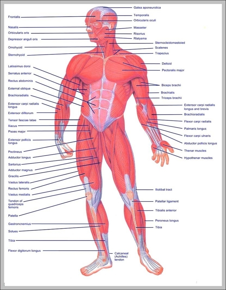
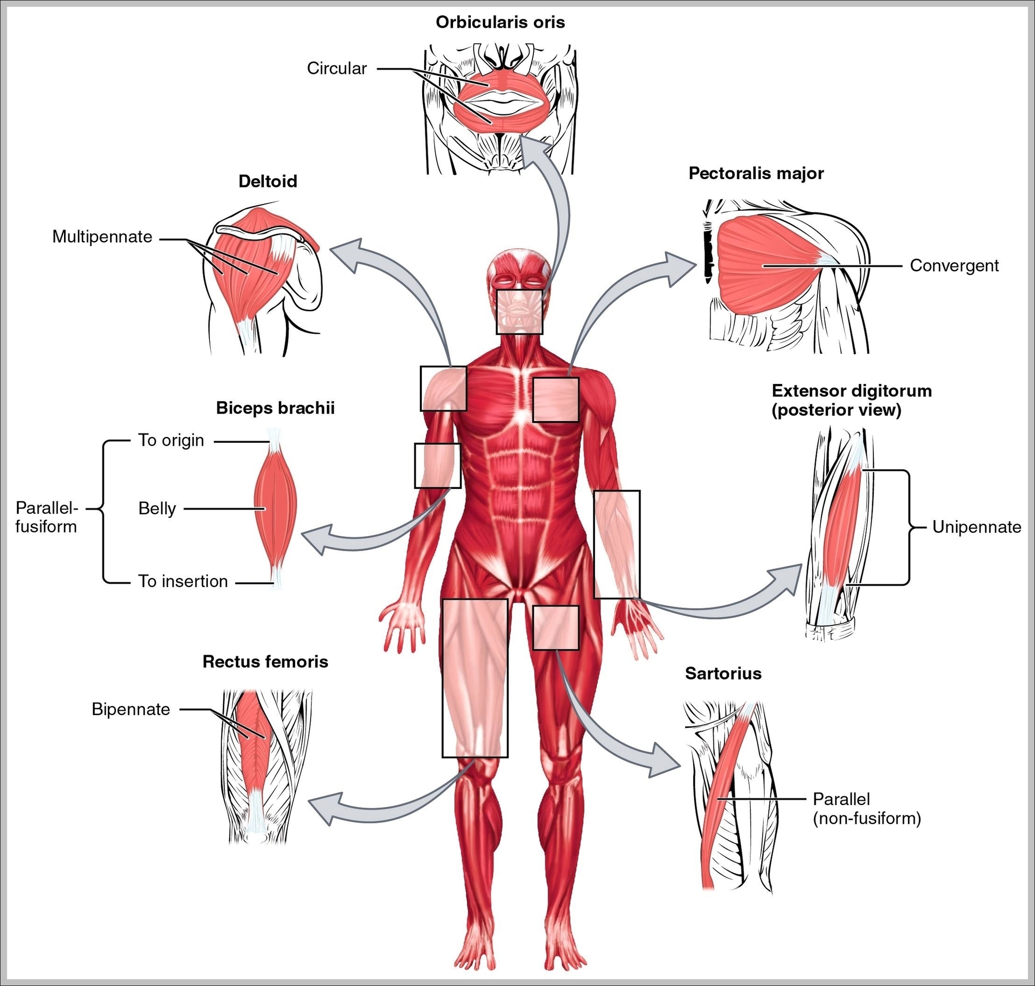
25,649 back muscle anatomy stock photos, vectors, and illustrations are available royalty-free.
25,649 back muscle anatomy stock photos, vectors, and illustrations are available royalty-free.