Posted inOrgans
Cardiac ConductionN
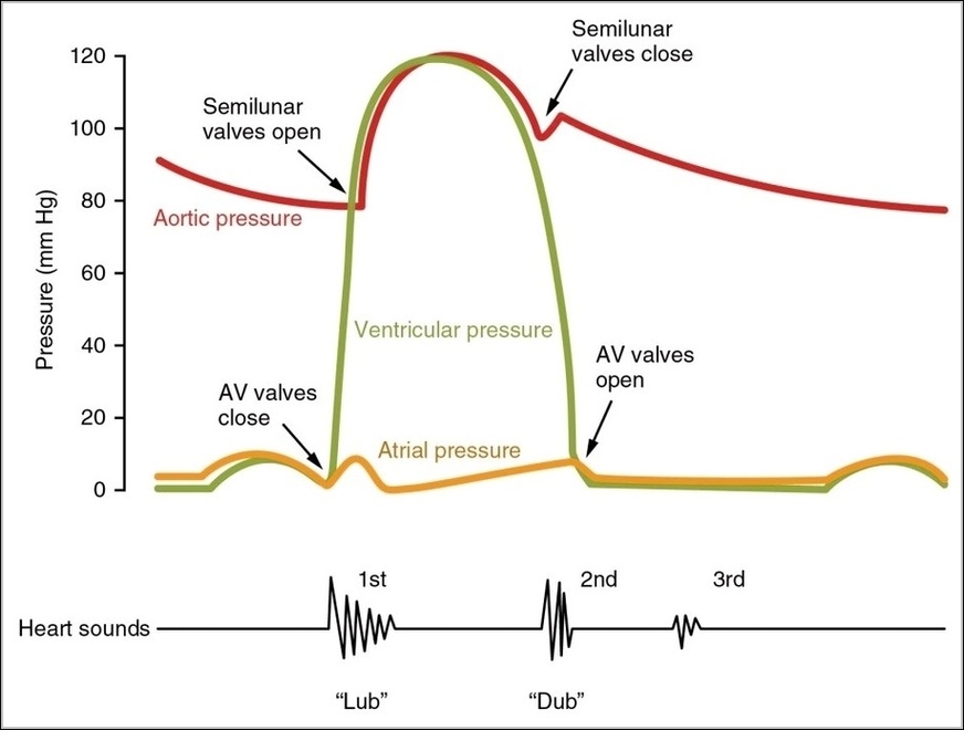
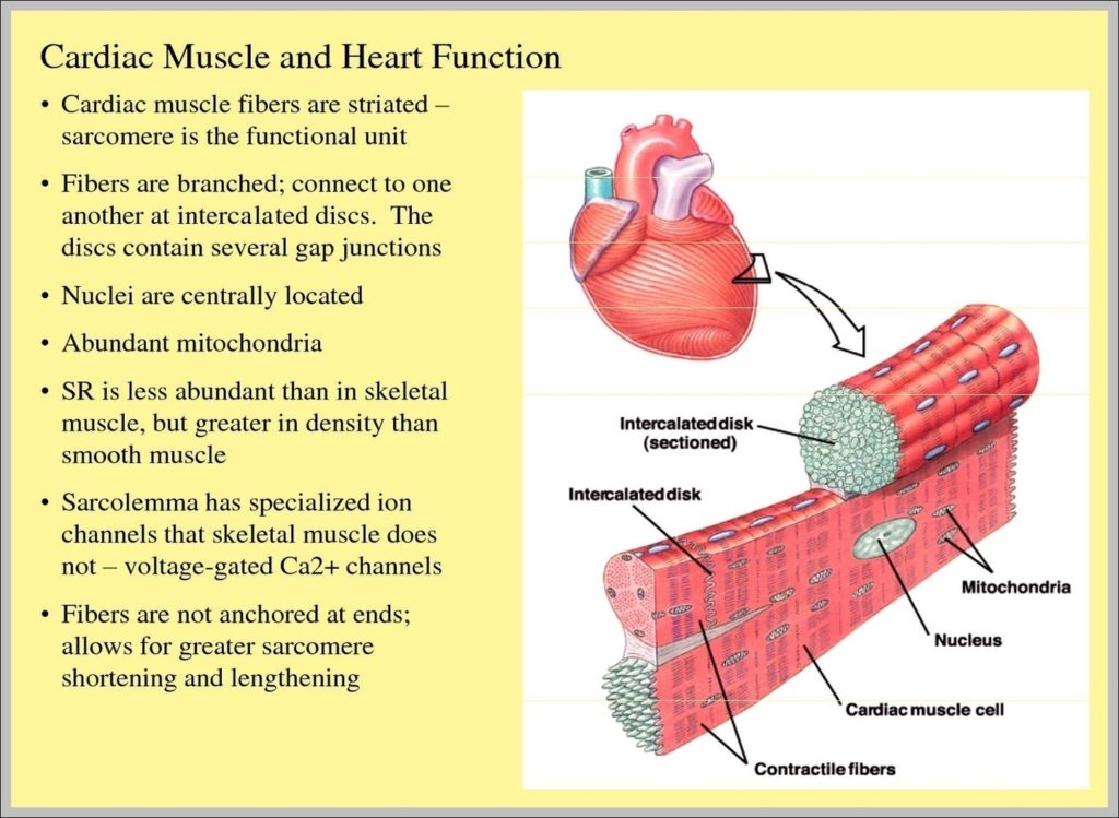
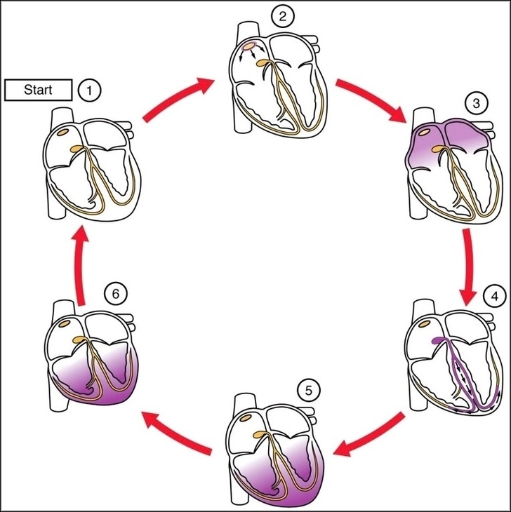
A cardiac conduction diagram maps the hearts electrical system from the sinoatrial node through the atrioventricular node and into the ventricles. It shows how timing and coordination ensure efficient contraction.…