Cardiac and pyloric sphincter are two sphincters of our stomach. They are specialized valves. The food remains inside the stomach due to the contraction of these two sphincters. What is the Difference Between Cardiac and Pyloric Sphincter?
Cardiac Sphincter: Location, Structure, and Function. The cardiac sphincter is a circular muscle located at the distal end of the esophagus. It relaxes to allow the passage of ingested food into the stomach, and constricts so that contents of stomach do not move back to the esophagus.
Cardiac Sphincter: Location, Structure, and Function. The cardiac sphincter is a circular muscle located at the distal end of the esophagus. It relaxes to allow the passage of ingested food into the stomach, and constricts so that contents of stomach do not move back to the esophagus.
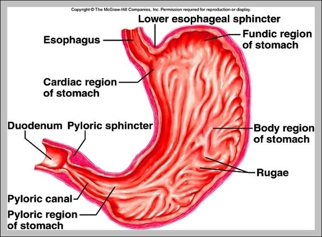
Cardiac And Pyloric Sphincter Image
Posted inDiagrams
Cardiac And Pyloric Sphincter Image
Post navigation
Previous Post
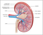 Function Of Kidney Image
Next Post
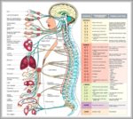
Parts Of The Body Diagram Image