Posted inMuscles
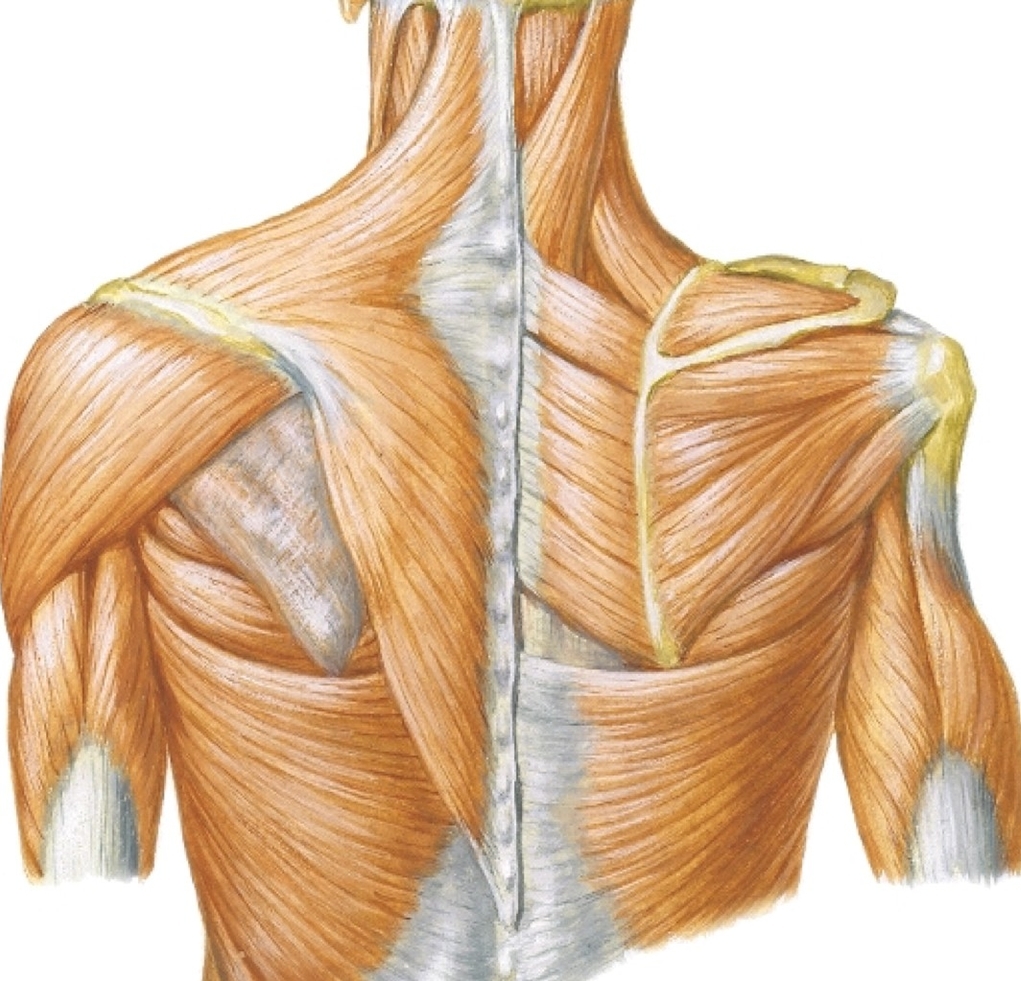
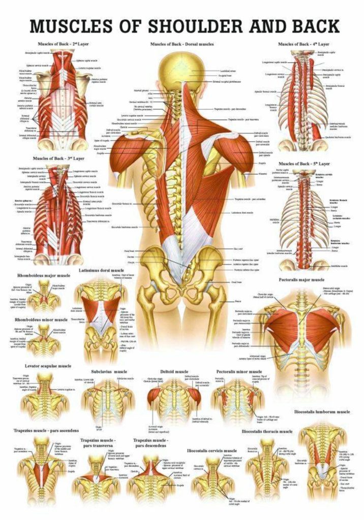
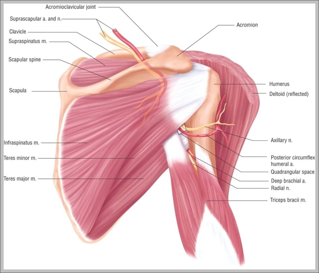
The Rotator Cuff Muscles of the Shoulder Joint Diagram
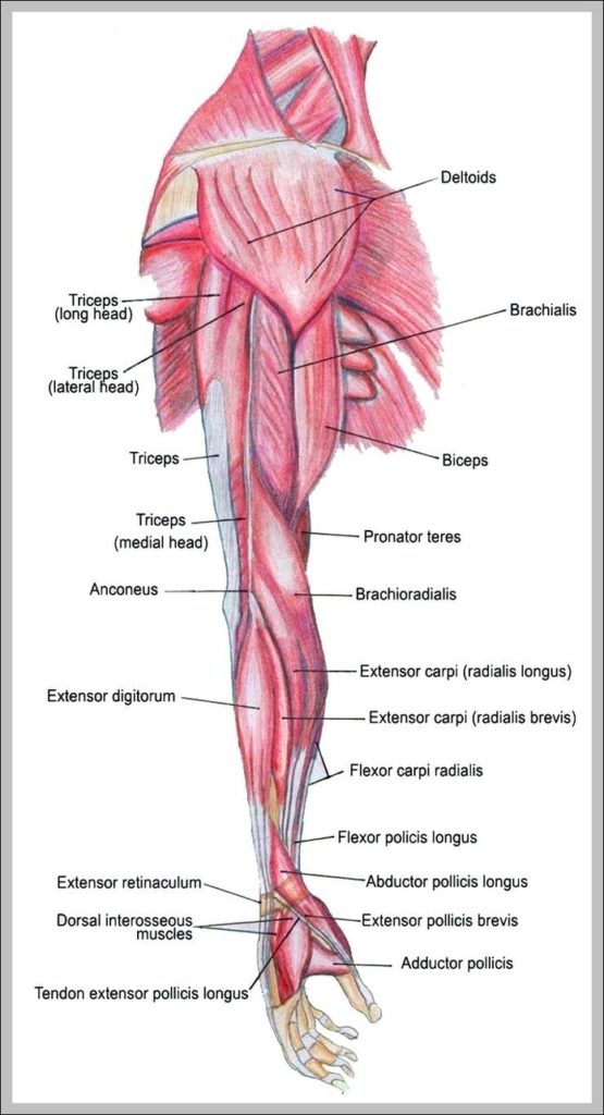
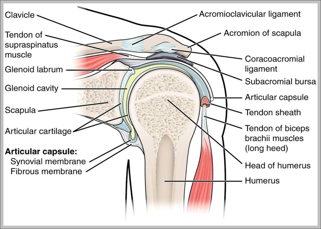
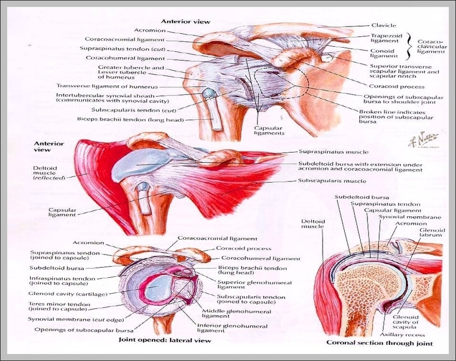
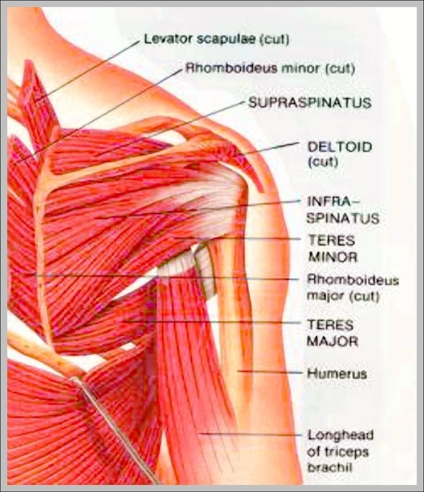
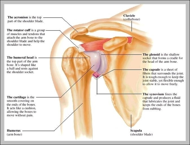
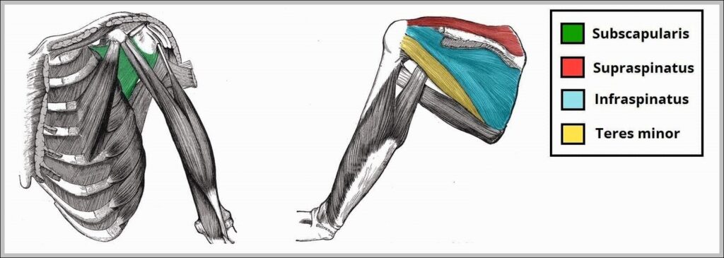
Rotator cuff muscles (supraspinatus, infraspinatus, teres minor, subscapularis) stabilize humeral head in glenoid during motion, with supraspinatus initiating abduction. Tears cause pain/weakness.