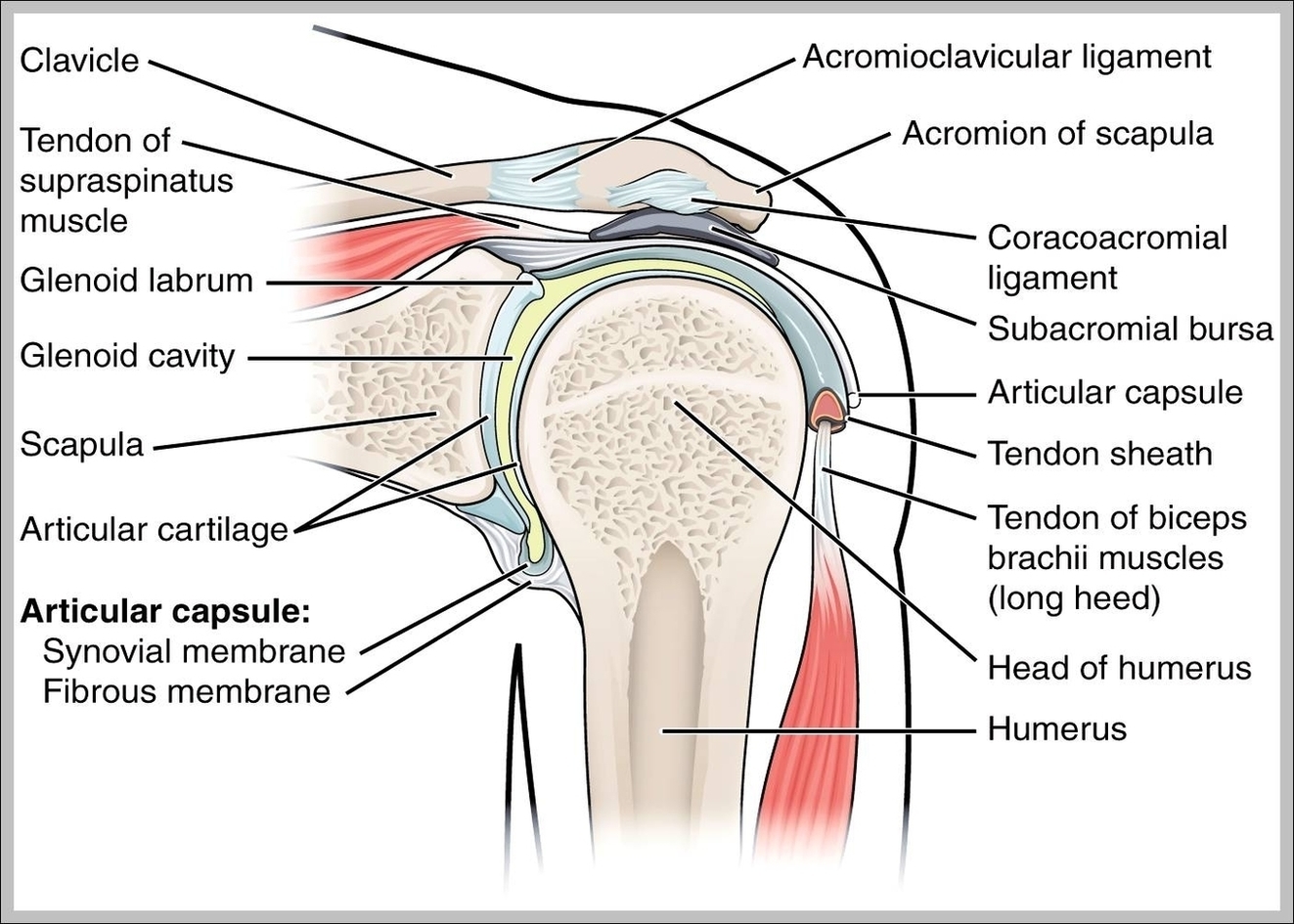
Shoulder Ligaments. Ligaments are soft tissue structures that connect bones to bones. There are several important ligaments in the shoulder. Glenohumeral Ligaments (GHL): A joint capsule is a watertight sac that surrounds a joint. In the shoulder, the joint capsule is formed by a group of ligaments that connect the humerus to the glenoid.
A joint capsule is a watertight sac that surrounds a joint. In the shoulder, the joint capsule is formed by a group of ligaments that connect the humerus to the glenoid. These ligaments are the main source of stability for the shoulder. They are the superior, middle and inferior glenohumeral ligaments.
A joint capsule is a watertight sac that surrounds a joint. In the shoulder, the joint capsule is formed by a group of ligaments that connect the humerus to the glenoid. These ligaments are the main source of stability for the shoulder. They are the superior, middle and inferior glenohumeral ligaments.
Ligaments Of The Shoulder Joint Image
Posted inDiagrams
Ligaments Of The Shoulder Joint Image
Post navigation
Previous Post
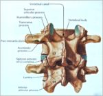 Pictures Of Vertebrae Image
Next Post
Ultrasound Degrees Image