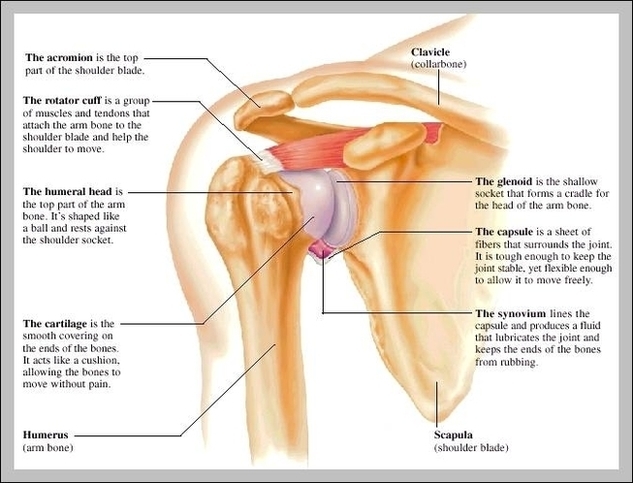
The shoulder joint is formed by the articulation of the head of the humerus with the glenoid cavity (or fossa) of the scapula. This gives rise to the alternate name for the shoulder joint – the glenohumeral joint.
The shoulder is one of the largest and most complex joints in the body. The shoulder joint is formed where the humerus (upper arm bone) fits into the scapula (shoulder blade), like a ball and socket. Other important bones in the shoulder include:
The Shoulder Capsule. The shoulder capsule surrounds the ball-and-socket part of the shoulder joint. The capsule separates the joint from the rest of the body and contains the joint fluid. Several ligaments make up parts of the joint capsule, and these ligaments are important in keeping the shoulder joint in proper position.

Shoulder Joint Name Image
Posted inDiagrams


