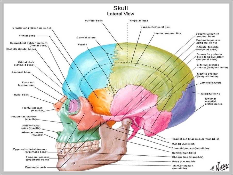
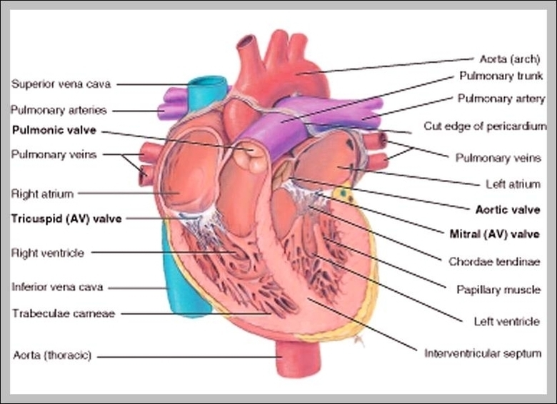
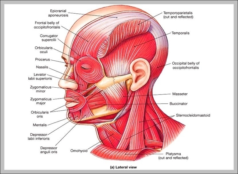
Skull Diagram Anatomy Image
Labeled Skull Diagram Overview image of an anterior view of the skull The idea behind using labeled diagrams is to get an overview of all of the structures within a given area. When it comes to testing your memory of these structures, previously having seen them altogether as a group should help you to remember them more easily.
The cranium (skull) is the skeletal structure of the head that supports the face and protects the brain. It is subdivided into the facial bones and the brain case.”Skull anatomy unlabeled In this image, you will find Skull anatomy in it.”
The cranium (skull) is the skeletal structure of the head that supports the face and protects the brain. It is subdivided into the facial bones and the brain case.”Skull anatomy unlabeled In this image, you will find Skull anatomy in it.”