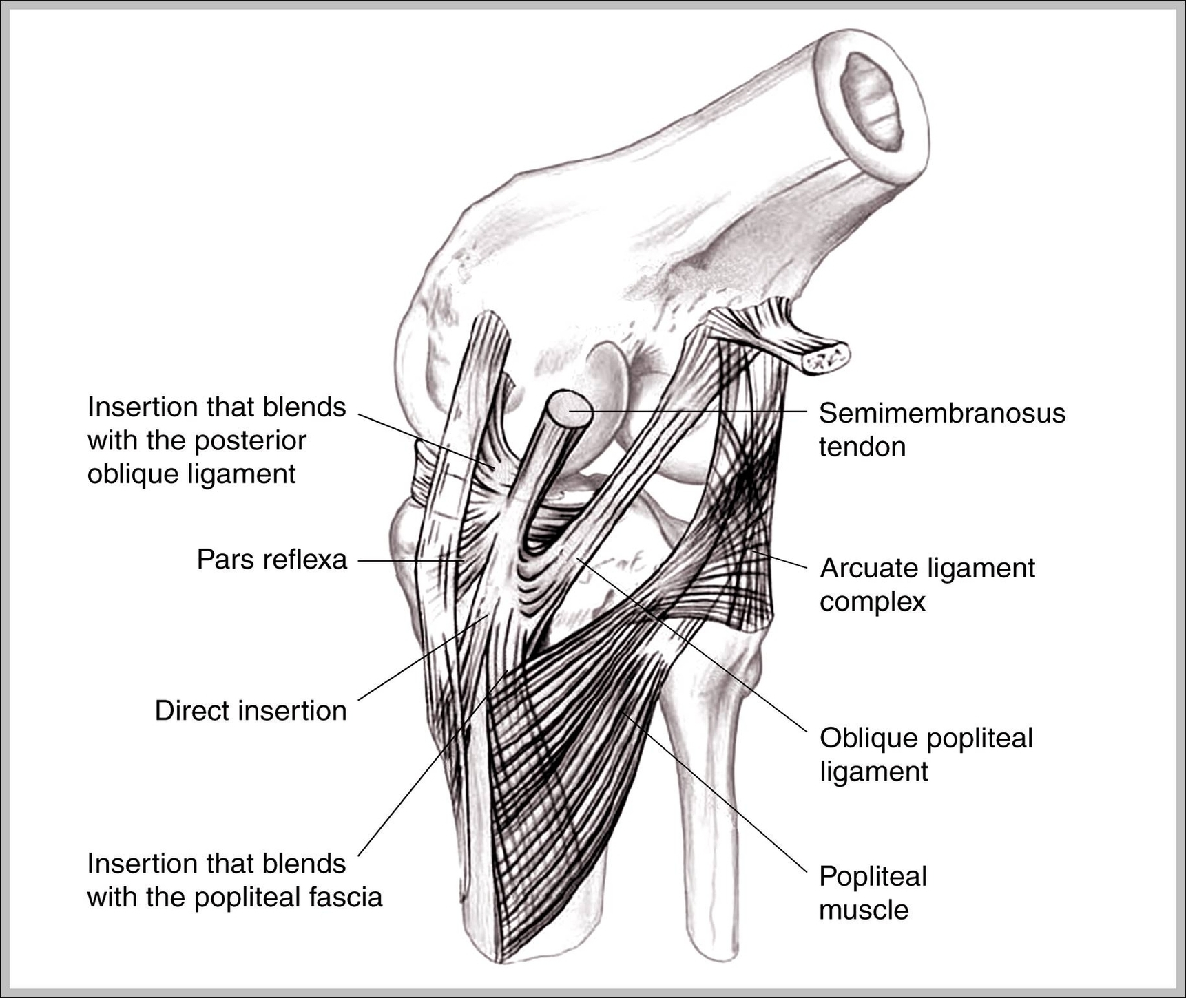
The medial collateral ligament ( MCL) of the knee is a flat, triangular band on its medial aspect that resists valgus forces. It forms part of the medial capsuloligamentous complex of the knee . The medial collateral ligament measures 8-10 cm in length and has superficial and deep portions 4.
MCL sprains are graded 1, 2 or 3 depending on severity. The medial collateral ligament (or MCL for short) connects the thigh bone (femur) to the shin bone (tibia) on the inside of your knee. It provides stability to your knee, preventing it from moving sideways.
MCL sprains are graded 1, 2 or 3 depending on severity. The medial collateral ligament (or MCL for short) connects the thigh bone (femur) to the shin bone (tibia) on the inside of your knee. It provides stability to your knee, preventing it from moving sideways.
Medial Collateral Ligament Image
Posted inDiagrams
Medial Collateral Ligament Image
Post navigation
Previous Post
 Picture Of Diaphragm Image
Next Post
External Body Parts Image