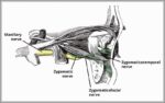
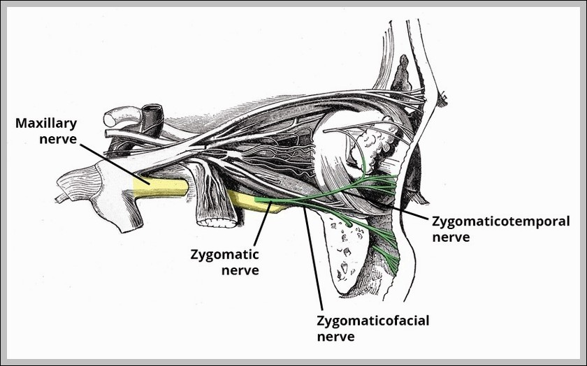
Kidney Image Image

3,867 human kidney stock photos and images available, or search for human kidney watercolors or human kidney anatomy to find more great stock photos and pictures.
Kidneys. The kidneys are bean-shaped organs that serve several essential regulatory roles in vertebrates. Their main function is to regulate the balance of Drone image, birds eye view about a blue swimming pool, with playing children. Aerial picture about a square shaped garden spot, with a larger kidney, and a

Kidney Image Image Diagram - Kidney Image Image Chart - Human anatomy diagrams and charts explained. This anatomy system diagram depicts Kidney Image Image with parts and labels. Best diagram to help learn about health, human body and medicine.

Kidney Image Image

Diagram Of The Body Systems Image

A human body diagram is a representation that outlines the major structures within the human body. This diagram should include a drawing or outline of the skeletal system and internal organs within the body.
Luckily, though, you can now download a free body diagram template and use it as a visual aid in the classroom. The students of today need understanding, so, you cannot teach a topic like this without a body diagram template.

Diagram Of The Body Systems Image Diagram - Diagram Of The Body Systems Image Chart - Human anatomy diagrams and charts explained. This anatomy system diagram depicts Diagram Of The Body Systems Image with parts and labels. Best diagram to help learn about health, human body and medicine.

Diagram Of The Body Systems Image

Diagram Of Human Organs In The Body Female Image

507 female body diagram stock photos and images available, or search for body silhouette or human body diagram to find more great stock photos and pictures. The Principal Glands Of The Female And Male Human Endocrine Systems.
7,751 organs of the human body diagram stock illustrations and vector graphics available royalty-free, or start a new search to explore more great stock images and vector art. internal organs and circulatory system Vector isolated illustration of human internal organs and circulatory system in man body.

Diagram Of Human Organs In The Body Female Image Diagram - Diagram Of Human Organs In The Body Female Image Chart - Human anatomy diagrams and charts explained. This anatomy system diagram depicts Diagram Of Human Organs In The Body Female Image with parts and labels. Best diagram to help learn about health, human body and medicine.

Diagram Of Human Organs In The Body Female Image

Images Of Skeleton Bones Image

398,454 skeleton bones stock photos, vectors, and illustrations are available royalty-free. See skeleton bones stock video clips
Skeleton in various poses is cartoon style Skeleton in various poses is cartoon style. Funny skeleton with various gestures. Vector illustration. human skeleton stock illustrations Skeleton in various poses is cartoon style.

Images Of Skeleton Bones Image Diagram - Images Of Skeleton Bones Image Chart - Human anatomy diagrams and charts explained. This anatomy system diagram depicts Images Of Skeleton Bones Image with parts and labels. Best diagram to help learn about health, human body and medicine.

Images Of Skeleton Bones Image

Body Anatomy Liver Image

4,742 human liver anatomy stock photos and images available, or start a new search to explore more stock photos and images.
The liver is a large, meaty organ that sits on the right side of the belly. Weighing about 3 pounds, the liver is reddish-brown in color and feels rubbery to the touch. Normally you can’t feel the liver, because it’s protected by the rib cage. The liver has two large sections, called the right and the left lobes.

Body Anatomy Liver Image Diagram - Body Anatomy Liver Image Chart - Human anatomy diagrams and charts explained. This anatomy system diagram depicts Body Anatomy Liver Image with parts and labels. Best diagram to help learn about health, human body and medicine.

Body Anatomy Liver Image

Arm Muscles Explained

Arm muscles. The arm muscles of the upper limb act on the elbow and shoulder joints to produce the various movements of the forearm. Five arm muscles play a role in these movements: biceps brachii, brachialis, coracobrachialis, triceps brachii and anconeus. The first three are primarily involved in forearm flexion, hence they are called flexors.
There are four muscles in you upper arm, which is delimited by your shoulder joint and your elbow joint. Three of them are located in the anterior compartment — the biceps brachii, brachialis, and coracobrachialis, while the forth is located in the posterior compartment — the triceps brachii). 1. The Muscles of the Biceps

Arm Muscles Explained Diagram - Arm Muscles Explained Chart - Human anatomy diagrams and charts explained. This anatomy system diagram depicts Arm Muscles Explained with parts and labels. Best diagram to help learn about health, human body and medicine.

Arm muscles explained

2nd Vertebrae Image

The second cervical vertebra of the spine is commonly called the C2 vertebra, axis, or epistropheus. A fracture of the C2 vertebra is known as a hangman’s fracture or axis vertebra fracture. A general term for a fracture of any of the seven bones in the neck (cervical vertebrae) is a broken neck.
L2 The L2 vertebra is the second lumbar spinal vertebra in the human body. Along with the other vertebrae, it makes up the spinal column, which supports the upper half of the human body. The lumbar…

2nd Vertebrae Image Diagram - 2nd Vertebrae Image Chart - Human anatomy diagrams and charts explained. This anatomy system diagram depicts 2nd Vertebrae Image with parts and labels. Best diagram to help learn about health, human body and medicine.

2nd Vertebrae Image

Labeled Muscle Diagram Image

98 muscular system labeled diagram stock photos and images available, or start a new search to explore more stock photos and images. Frontal view of the muscular system of the male human body with descriptive labels pointing to the muscles on a white background. Labeled Anatomy Chart of Neck and Shoulder Muscles on White…
Labeled human anatomy diagram of man’s full body muscular system from a posterior view on a white background. Labeled chart of the facial muscles of a woman, frontal view on a white background. Male Shoulder Ligaments and Biceps Muscles Isolated in Skeleton…

Labeled Muscle Diagram Image Diagram - Labeled Muscle Diagram Image Chart - Human anatomy diagrams and charts explained. This anatomy system diagram depicts Labeled Muscle Diagram Image with parts and labels. Best diagram to help learn about health, human body and medicine.

Labeled Muscle Diagram Image

Hand Bones Pictures Image

Hand Bones Pictures Image Diagram - Hand Bones Pictures Image Chart - Human anatomy diagrams and charts explained. This anatomy system diagram depicts Hand Bones Pictures Image with parts and labels. Best diagram to help learn about health, human body and medicine.

Hand Bones Pictures Image

Pictures Of The Human Spine Image

Pictures Of The Human Spine Image Diagram - Pictures Of The Human Spine Image Chart - Human anatomy diagrams and charts explained. This anatomy system diagram depicts Pictures Of The Human Spine Image with parts and labels. Best diagram to help learn about health, human body and medicine.

Pictures Of The Human Spine Image

Pancreas Functions Image

Human Nervous System. The central nervous system (CNS) together with the peripheral nervous system (PNS) together constitute the human neural system. CNS consists of the control and information processing units, the brain and the spinal cord. PNS consists of the nerves that connect peripheral parts to the brain and the spinal cord viz. the CNS.
One of the most complex organ system to ever evolve, the human nervous system consists of two parts, namely: Central Nervous System (consists of the brain and spinal cord) Peripheral Nervous System (includes all the nerves of the body)

Pancreas Functions Image Diagram - Pancreas Functions Image Chart - Human anatomy diagrams and charts explained. This anatomy system diagram depicts Pancreas Functions Image with parts and labels. Best diagram to help learn about health, human body and medicine.

Pancreas Functions Image

Sacral Spine Image

The strong muscles that move the ankle originate in the leg. The most well-known are the gastrocnemius and soleus muscles, which are the strong calf muscles that allow you to push up on your toes.
The anatomy of the foot is incredibly complex. This introduction to the anatomy of the foot and ankle will be very general and highlight the most relevant structures. The ankle joint or tibiotalar joint is formed where the top of the talus (the uppermost bone in the foot) and the tibia (shin bone) and fibula meet.

Sacral Spine Image Diagram - Sacral Spine Image Chart - Human anatomy diagrams and charts explained. This anatomy system diagram depicts Sacral Spine Image with parts and labels. Best diagram to help learn about health, human body and medicine.

Sacral Spine Image

L Spine Image

Your back consists of a complex array of bones, discs, nerves, joints, and muscles. The muscles of your back support your spine, attach your pelvis and shoulders to your trunk, and provide mobility and stability to your trunk and spine. The anatomy of your back muscles can be complex.
25,649 back muscle anatomy stock photos, vectors, and illustrations are available royalty-free.

L Spine Image Diagram - L Spine Image Chart - Human anatomy diagrams and charts explained. This anatomy system diagram depicts L Spine Image with parts and labels. Best diagram to help learn about health, human body and medicine.

L Spine Image

Hip And Thigh Muscles Image

The hip muscles encompass many muscles of the hip and thigh whose main function is to act on the thigh at the hip joint and stabilize the pelvis. Without them, walking would be impossible. They can be divided into three main groups:
See something you like? Save 25% when you use code SHARE25. 6,153 anatomy hip muscles stock photos, vectors, and illustrations are available royalty-free.

Hip And Thigh Muscles Image Diagram - Hip And Thigh Muscles Image Chart - Human anatomy diagrams and charts explained. This anatomy system diagram depicts Hip And Thigh Muscles Image with parts and labels. Best diagram to help learn about health, human body and medicine.

Hip And Thigh Muscles Image

Adenoids And Tonsils Image

Adenoids and tonsils Stock Photo Images. 172 Adenoids and tonsils royalty free images and photography available to buy from thousands of stock photographers. Female ent doctor examining patient’s mouth at modern clinic.
Lingual tonsils are at base of tongue directly below endotracheal tube at point where tongue meets epiglottis. Adenoids are visualized via degree endoscope. Adenoids lie in nasopharynx, lined laterally by torus tubarius.Enlarged AdenoidsRed Blood Spots On Tonsils Tonsils Diagram Adenoids – Grade Revus-Project

Adenoids And Tonsils Image Diagram - Adenoids And Tonsils Image Chart - Human anatomy diagrams and charts explained. This anatomy system diagram depicts Adenoids And Tonsils Image with parts and labels. Best diagram to help learn about health, human body and medicine.

Adenoids And Tonsils Image

Blood Supply To Small Intestine Image

Blood Supply and Innervation of the Small Intestine. The neurovascular supply of any area of the body is the network in which the blood is pumped and the structures are innervated.
The blood supply to the small and large bowel is derived from the celiac artery and SMA. The celiac axis primarily provides blood flow to the stomach, liver, spleen, and pancreas but is also a source of collateral flow when blood flow in the SMA is reduced.

Blood Supply To Small Intestine Image Diagram - Blood Supply To Small Intestine Image Chart - Human anatomy diagrams and charts explained. This anatomy system diagram depicts Blood Supply To Small Intestine Image with parts and labels. Best diagram to help learn about health, human body and medicine.

Blood Supply To Small Intestine Image

Nerves Of The Neck Image

Upper Back Muscles. The deltoid, teres major, teres minor, infraspinatus, supraspinatus (not shown) and subscapularis muscles (not shown) all extend from the scapula to the humerus and act on the shoulder joint.
25,649 back muscle anatomy stock photos, vectors, and illustrations are available royalty-free.

Nerves Of The Neck Image Diagram - Nerves Of The Neck Image Chart - Human anatomy diagrams and charts explained. This anatomy system diagram depicts Nerves Of The Neck Image with parts and labels. Best diagram to help learn about health, human body and medicine.

Nerves Of The Neck Image

Pancreas Anatomy Image

Your spleen is a small organ that sits inside your left rib cage, just above your stomach. In adults, the spleen is about the size of an avocado. The spleen is part of your lymphatic system (which is part of your immune system ).
3,264 spleen stock photos and images available, or search for spleen icon or enlarged spleen to find more great stock photos and pictures.

Pancreas Anatomy Image Diagram - Pancreas Anatomy Image Chart - Human anatomy diagrams and charts explained. This anatomy system diagram depicts Pancreas Anatomy Image with parts and labels. Best diagram to help learn about health, human body and medicine.

Pancreas Anatomy Image

Anatomy Of The Shoulder Joint Image

shoulder joint anatomy shoulder joint anatomy. Bones (Scapula, Humerus, Coracoid process, Acromion , Muscle (Biceps, Supraspinatus) and ligament (Coracohumeral) shoulder anatomy stock illustrations shoulder joint anatomy.
13,987 shoulder anatomy stock photos and images available, or search for shoulder anatomy muscles to find more great stock photos and pictures. Man with extended arm. Illustrated representation of the structure and musculature of the human arm. Studio man portrait. shoulder anatomy stock pictures, royalty-free photos & images

Anatomy Of The Shoulder Joint Image Diagram - Anatomy Of The Shoulder Joint Image Chart - Human anatomy diagrams and charts explained. This anatomy system diagram depicts Anatomy Of The Shoulder Joint Image with parts and labels. Best diagram to help learn about health, human body and medicine.

Anatomy Of The Shoulder Joint Image

Tendons In The Knee Diagram Image

The knee is composed of bones, tendons, ligaments, muscles, cartilage, and other soft tissues. Therefore, t his article will focus on the tendons of the knee and injuries to the tendons of the knee. Tendons are structures that connect muscles to bones, and many muscles cross the knee joint, which help maintain its structure and stability.
To understand one of the most complex joints of our body i.e. the knee joint, you need a perfectly labeled diagram of the knee. This will help you to understand the mechanism as well as the working.

Tendons In The Knee Diagram Image Diagram - Tendons In The Knee Diagram Image Chart - Human anatomy diagrams and charts explained. This anatomy system diagram depicts Tendons In The Knee Diagram Image with parts and labels. Best diagram to help learn about health, human body and medicine.

Tendons In The Knee Diagram Image