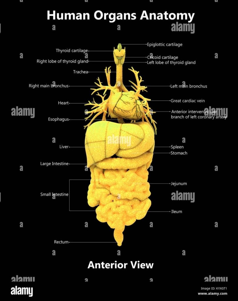


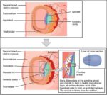

Posted inOrgans
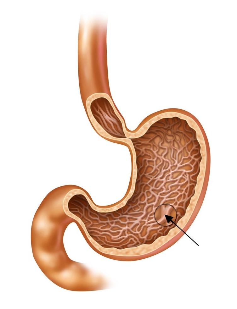
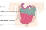
Small IntestineN
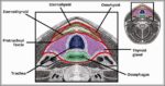
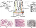
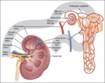
Small intestine is coiled 6-7 meter tube from pylorus to ileocecal valve divided duodenum C-shaped around pancreas head receiving bile pancreatic ducts, jejunum upper two-fifths major absorption site, ileum lower…