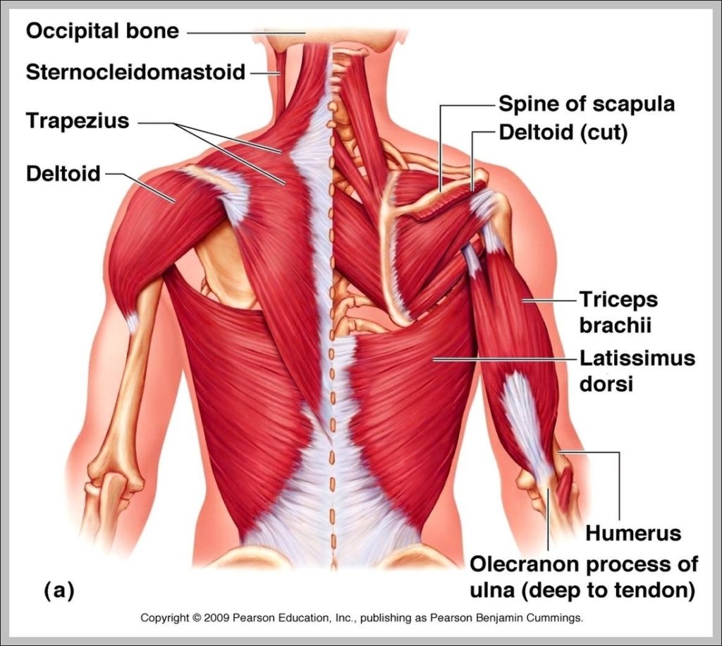
The deltoid is a thick, triangular shoulder muscle. It gets its name because of its similar shape to the Greek letter ‘delta’ (Δ). The muscle has a wide origin spanning…
25,649 back muscle anatomy stock photos, vectors, and illustrations are available royalty-free. Best viewed on 1280 x 768 px resolution in any modern browser. This article is about Pictures Of…
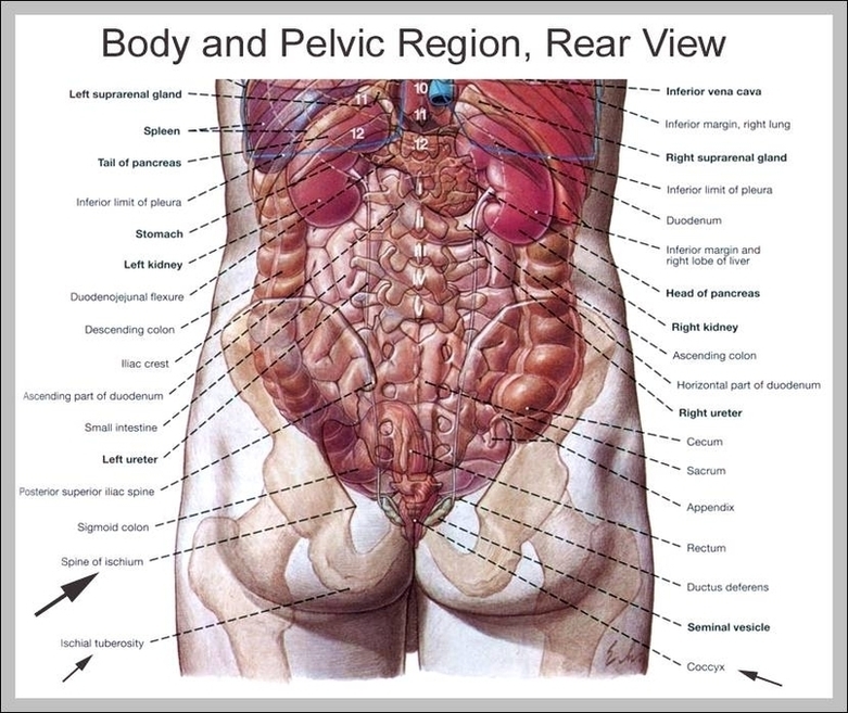
223 lower back skeleton stock photos and images available, or start a new search to explore more stock photos and images. The urinary system includes several structures, the kidneys, the…
3,344 knee anatomy stock photos and images available, or search for knee anatomy illustration or human knee anatomy to find more great stock photos and pictures. Picture of the Knee.…
52,129 human back anatomy stock photos, vectors, and illustrations are available royalty-free. Human anatomy xray view of intestines, on light back. Human anatomy xray view of intestines, showing stomach, colon,…
25,649 back muscle anatomy stock photos, vectors, and illustrations are available royalty-free. Best viewed on 1280 x 768 px resolution in any modern browser. This article is about Pictures Of…
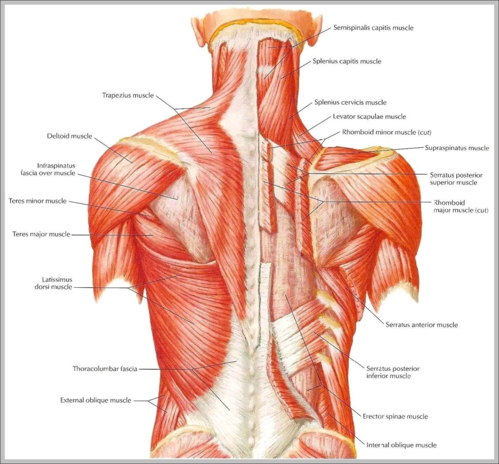
Common upper back pain symptoms include muscle stiffness, tightness, and tenderness that may affect the shoulders and neck. Upper back pain is often related to muscle or soft tissue problems,…
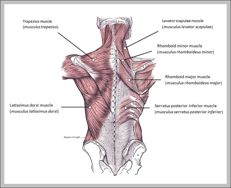
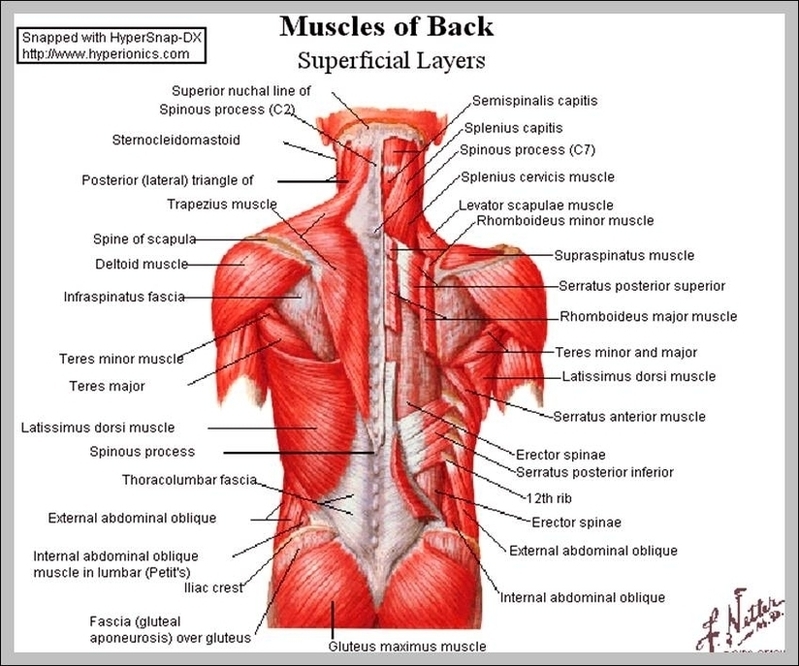
25,649 back muscle anatomy stock photos, vectors, and illustrations are available royalty-free. As with other parts of the body, the back has several layers of muscles. Some are closer to…
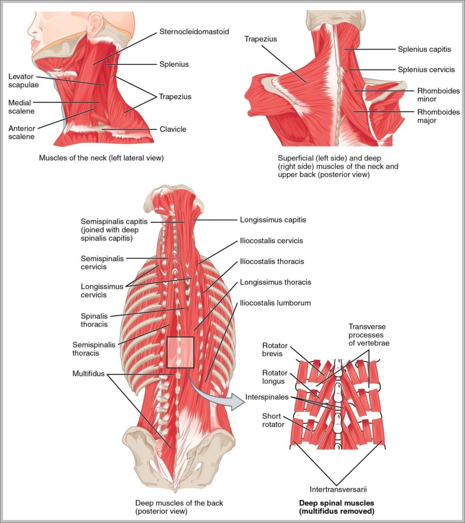
Muscles of neck. Neck muscles are bodies of tissue that produce motion in the neck when stimulated. The muscles of the neck run from the base of the skull to…
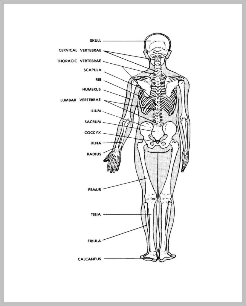
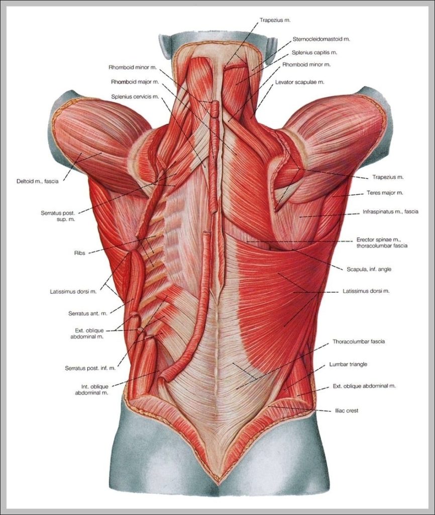
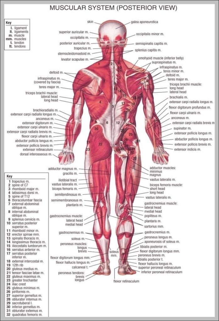
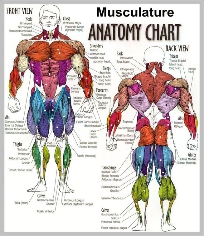
Your back consists of a complex array of bones, discs, nerves, joints, and muscles. The muscles of your back support your spine, attach your pelvis and shoulders to your trunk,…
Lower back muscle anatomy includes the Multifidus, Longissimus, Spinalis, and Quadratus Lumborum. The muscles of the low back work together with the transverse abdominal muscles to increase intra-abdominal pressure. The…
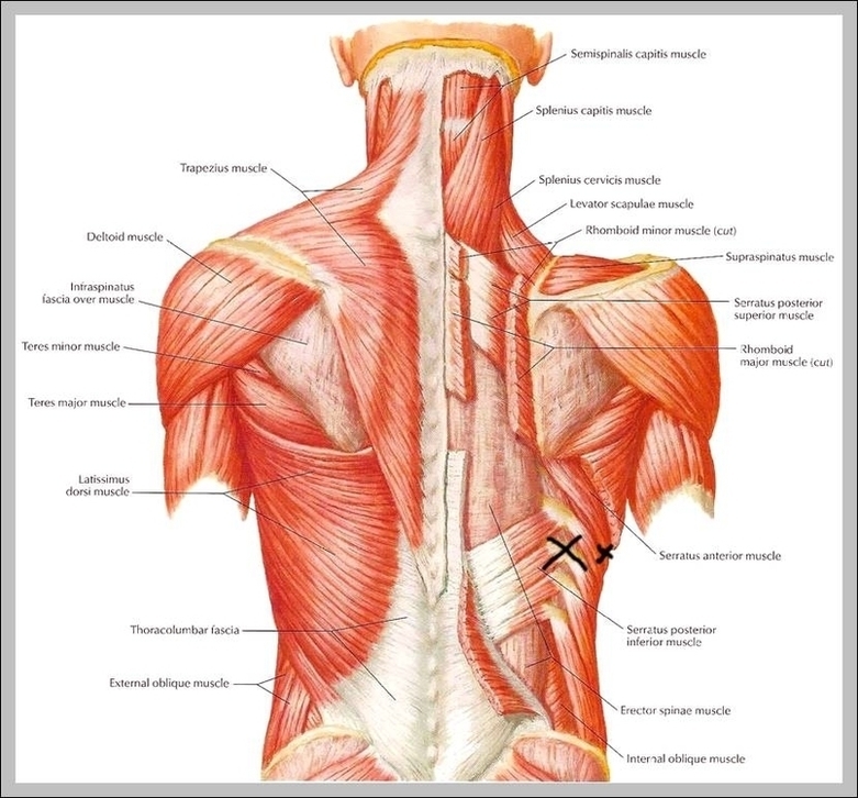
Upper Back Muscles. The deltoid, teres major, teres minor, infraspinatus, supraspinatus (not shown) and subscapularis muscles (not shown) all extend from the scapula to the humerus and act on the…
Your lower back (lumbar spine) is the anatomic region between your lowest rib and the upper part of the buttock. 1 Your spine in this region has a natural inward…
25,649 back muscle anatomy stock photos, vectors, and illustrations are available royalty-free. As with other parts of the body, the back has several layers of muscles. Some are closer to…
Your back consists of a complex array of bones, discs, nerves, joints, and muscles. The muscles of your back support your spine, attach your pelvis and shoulders to your trunk,…
52,129 human back anatomy stock photos, vectors, and illustrations are available royalty-free. 7,751 organs of the human body diagram stock illustrations and vector graphics available royalty-free, or start a new…
Your back consists of a complex array of bones, discs, nerves, joints, and muscles. The muscles of your back support your spine, attach your pelvis and shoulders to your trunk,…
25,649 back muscle anatomy stock photos, vectors, and illustrations are available royalty-free. + Custom ... Male muscle anatomy of the human back. Paired bone, triangular in shape, articulating with the…