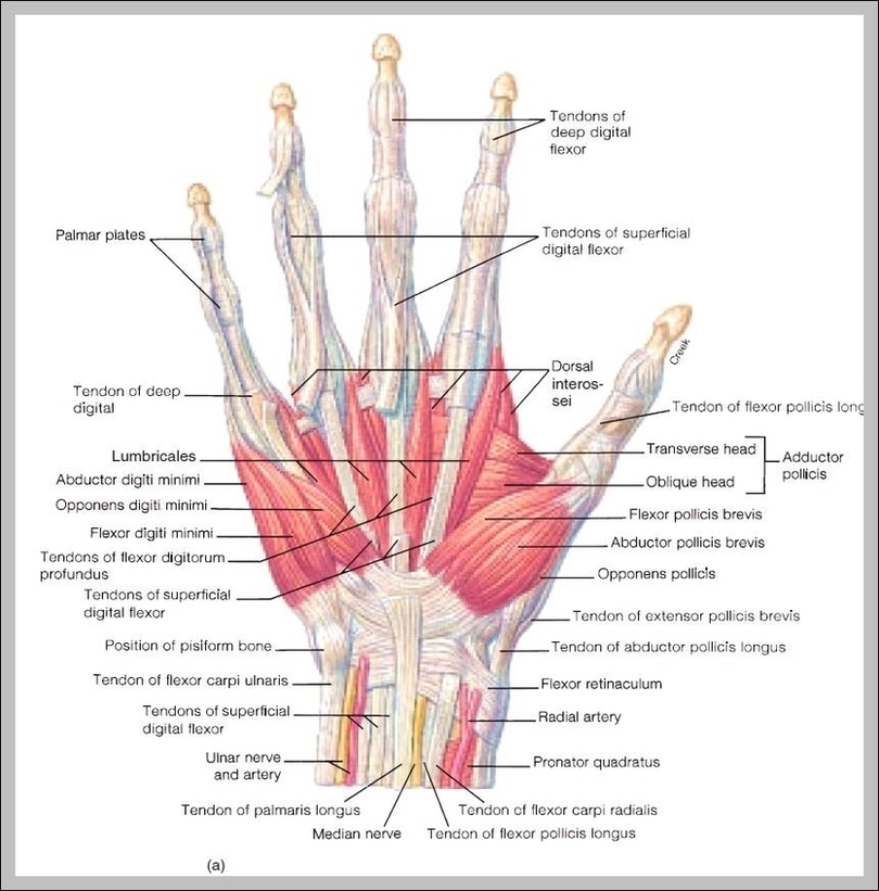
Fingers have a complex anatomy. Each finger has 3 phalanges (bones) and 3 hinged joints; the thumb has two of each. Ligaments connect finger bones and help keep them in place. Tendons connect muscles to bones. Finger movement is controlled by muscles in the forearms that pull on finger tendons.
Fingers are constructed of ligaments (strong supportive tissue connecting bone to bone), tendons (attachment tissue from muscle to bone), and three phalanges (bones). There are no muscles in the fingers; and fingers move by the pull of forearm muscles on the tendons.
Fingers are constructed of ligaments (strong supportive tissue connecting bone to bone), tendons (attachment tissue from muscle to bone), and three phalanges (bones). There are no muscles in the fingers; and fingers move by the pull of forearm muscles on the tendons.
Finger Anatomy Image
Posted inDiagrams
Finger Anatomy Image
Post navigation
Previous Post
 Human Body Internal Organs Diagram Image