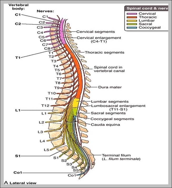
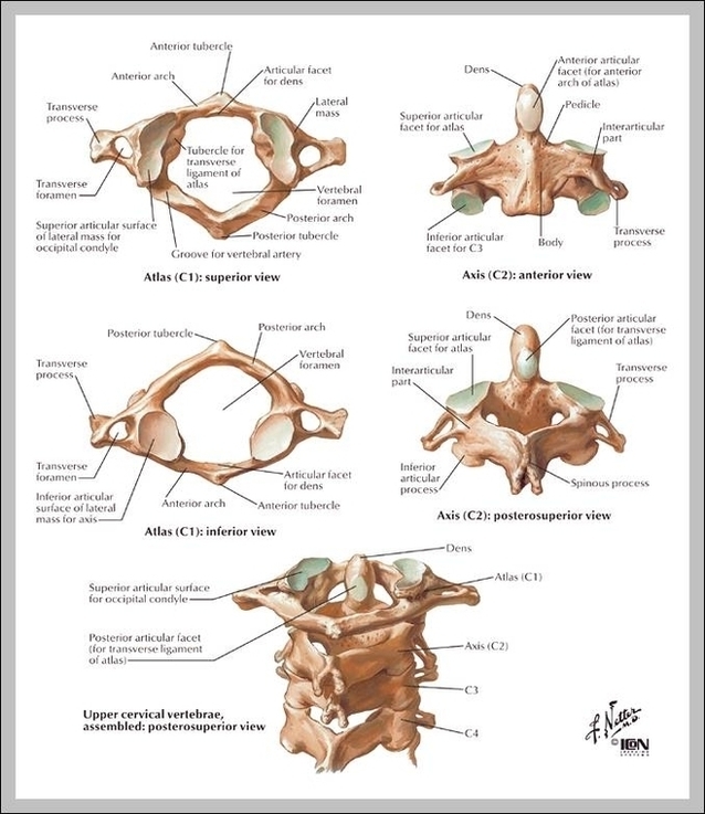
T 11 Vertebrae Image
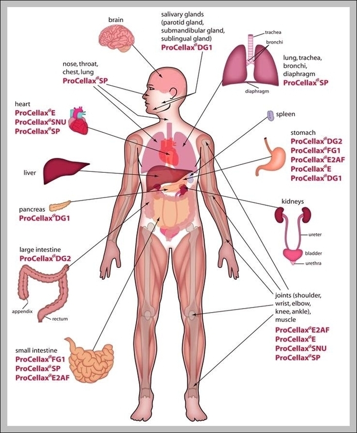
Anatomy of male muscles in upper body, anterior view. This image shows a cross section of the trachea and the lungs, in their places in the upper body. Upper body, Human male chest internal organs lung heart and stomach part model figure for medical education. Photo of a male neck. Facial hair and body hair can be seen.
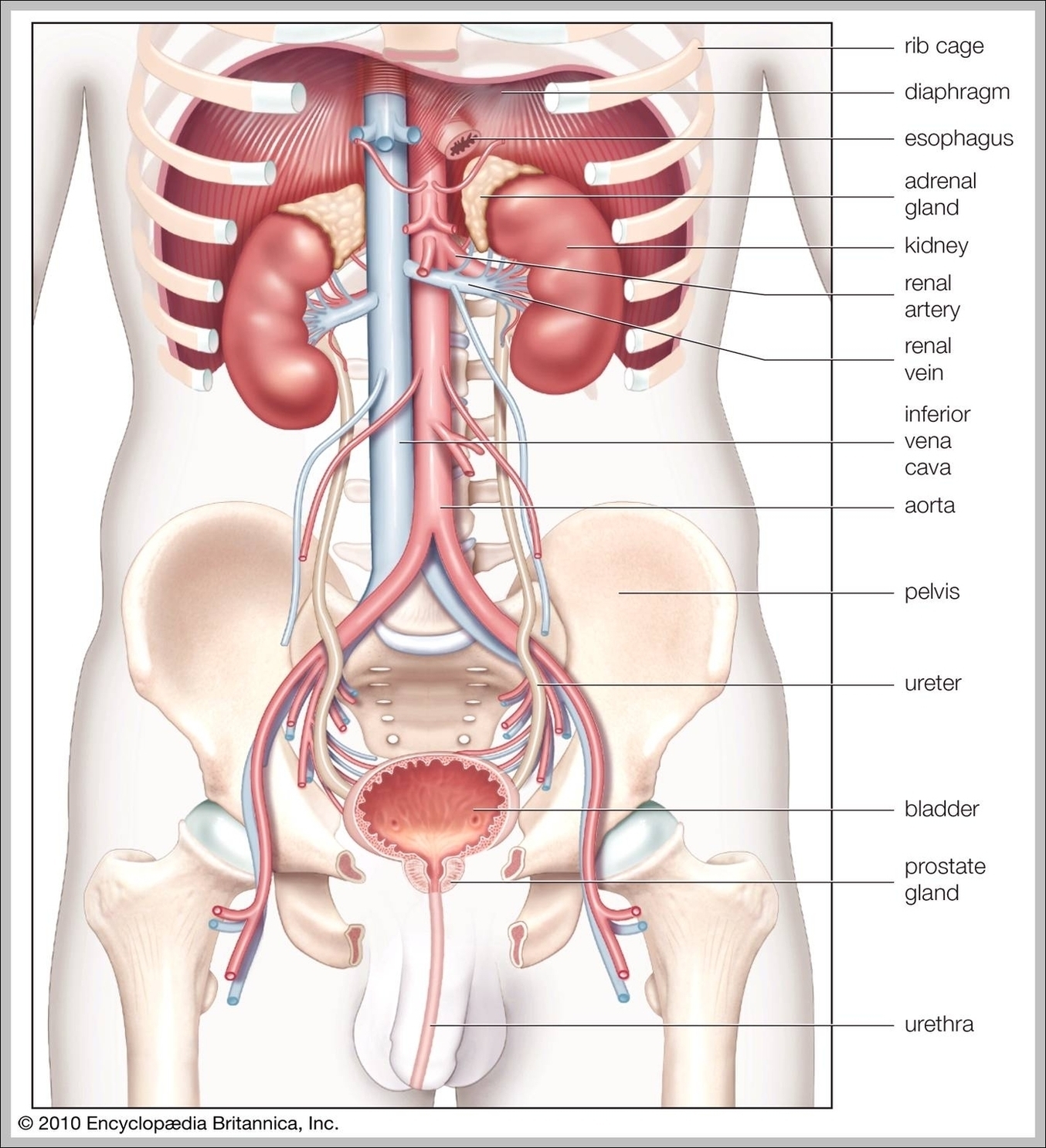
Anatomy of male muscles in upper body, posterior view. This image shows the upper organs of the digestive system. Angled view of the upper body of the female muscular anatomy. Very educational. Angled view of the upper body of the female muscular anatomy.
Anatomy of male muscles in upper body, posterior view. This image shows the upper organs of the digestive system. Angled view of the upper body of the female muscular anatomy. Very educational. Angled view of the upper body of the female muscular anatomy.