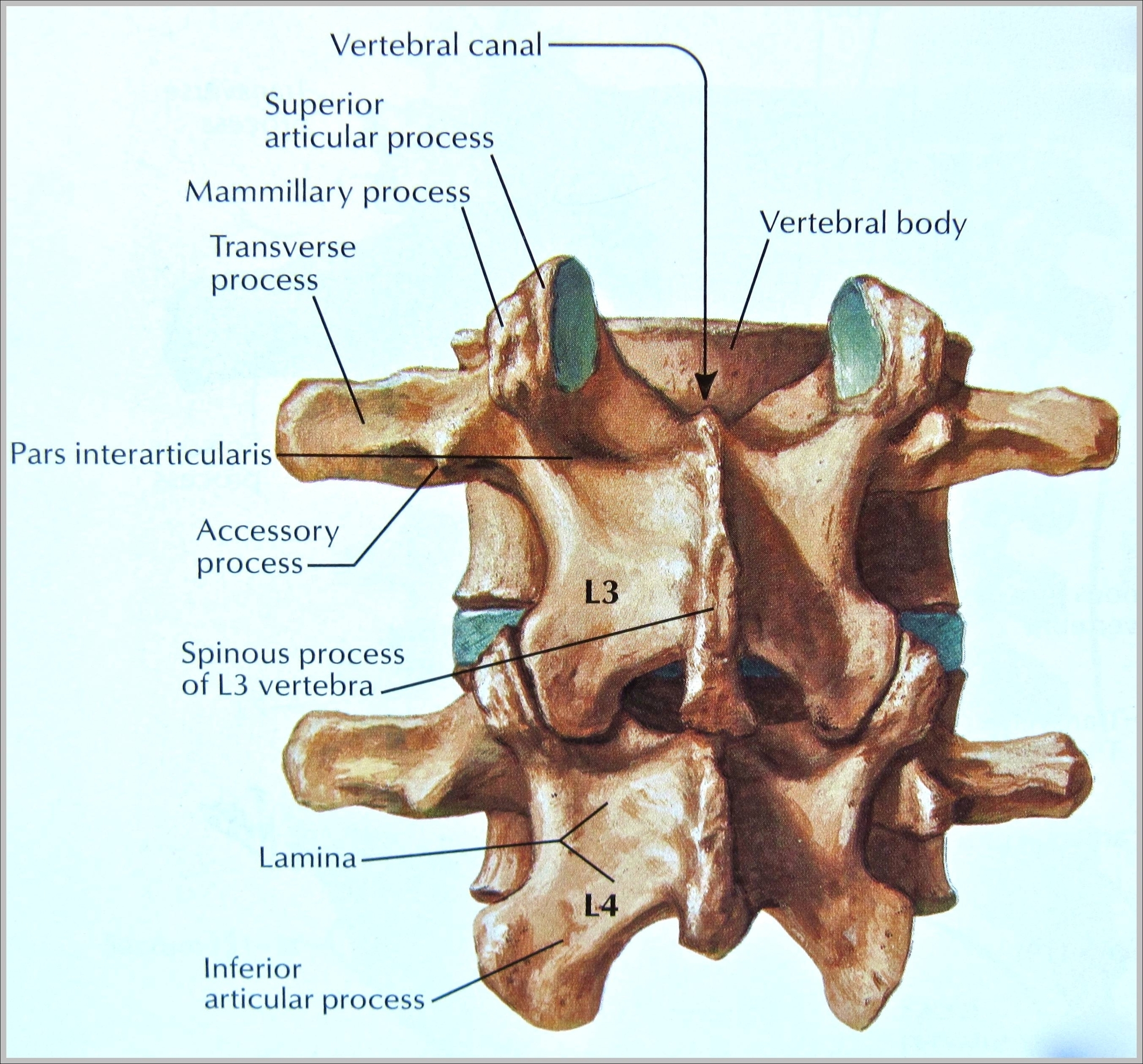
Five (or in some cases, six) vertebrae make up the lumbar spine, which provides support for much of the upper body and is rather flexible. The third lumbar spine vertebra (L3) is located in the middle of the lumbar spine, making it particularly susceptible to wear and tear. It is one of the most common sites for causes of chronic lower back pain.
The L3 vertebra is in the middle of the five lumbar vertebrae in the lower back portion of the spinal column. The L3 vertebra, or third lumbar vertebra, is one of the most common sites for the occurrence of a herniated disc and other spinal conditions that can cause chronic lower back pain.
The L3 vertebra is in the middle of the five lumbar vertebrae in the lower back portion of the spinal column. The L3 vertebra, or third lumbar vertebra, is one of the most common sites for the occurrence of a herniated disc and other spinal conditions that can cause chronic lower back pain.
3rd Vertebrae Image
Posted inDiagrams
3rd Vertebrae Image
Post navigation
Previous Post
 Spine Bone Image
Next Post
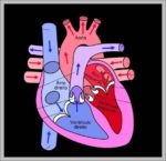
Images Of The Human Heart Image