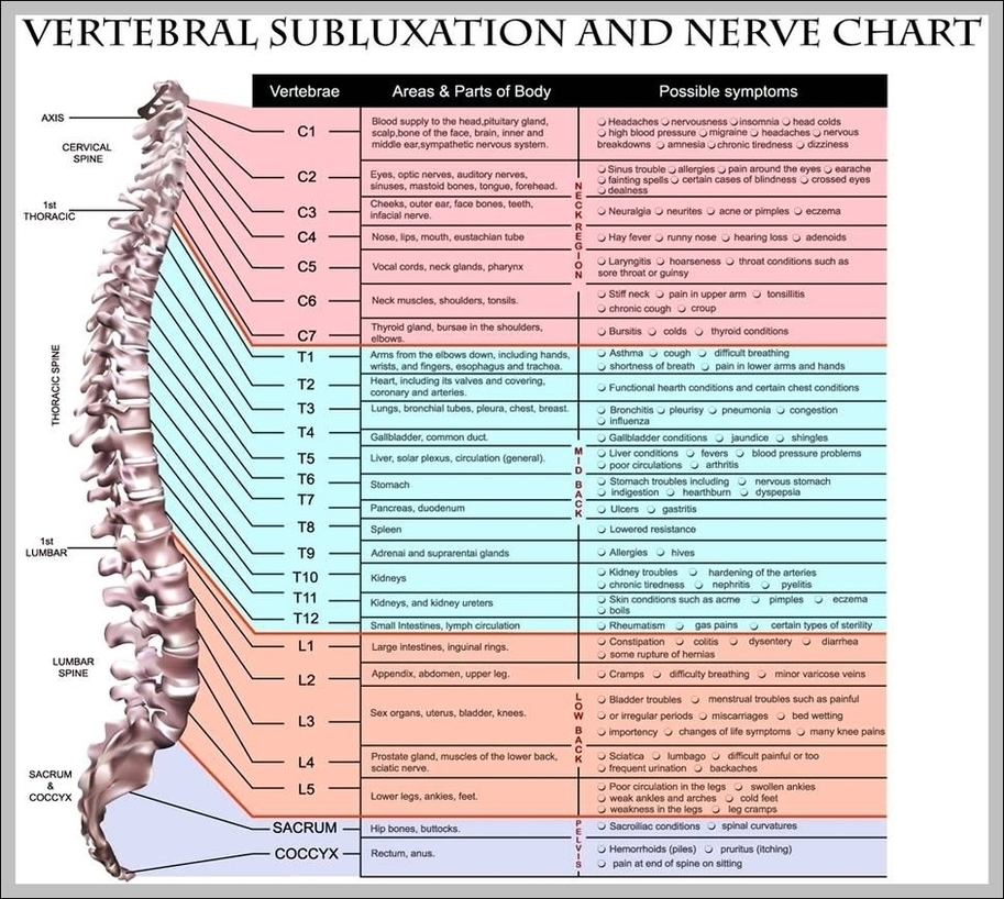
How To Use The Spinal Nerve Chart: On the chart below you will see 4 Columns (Vertebral Level, Nerve Root, Innervation, and Possible Symptoms). Under ‘Vertebral Level’: Simply line up the “Vertebral Level” with the “Possible Symptoms” and you will see some surprising connections of symptoms that relate to your spine.
Fig 1. Lateral labeled diagram of the human vertebral spinal column showing vertebrae numbering order and the 5 different regions of the spine. The Atlas is the topmost vertebra, and along with C2, forms the joint connecting the skull and spine.
Fig 1. Lateral labeled diagram of the human vertebral spinal column showing vertebrae numbering order and the 5 different regions of the spine. The Atlas is the topmost vertebra, and along with C2, forms the joint connecting the skull and spine.
Spinal Vertebrae Chart Image
Posted inDiagrams
Spinal Vertebrae Chart Image
Post navigation
Previous Post
Next Post
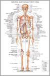
Anatomical Chart Image