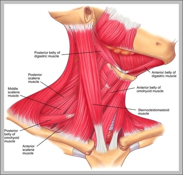
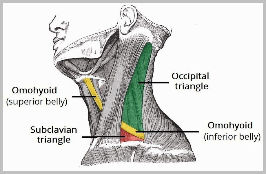
Head And Neck Muscles Example Of

The muscles of the head and neck are intricate and diverse, responsible for a variety of crucial tasks such as movement of the head and neck, chewing and swallowing, speech, facial expressions, and movement of the eyes. These tasks require both strong, forceful movements and some of the fastest, finest, and most delicate adjustments in the entire human body.

Facial Muscles

The muscles of the face are unique among groups of muscles in the body. While most muscles connect to and move only bones, facial muscles mostly connect bones to skin. These muscles, including the zygomaticus major and orbicularis oris, pull on the skin to produce a seemingly infinite number of facial expressions and to move the lips and cheeks during speech and eating.

Muscles of Mastication

Producing the body’s ability to close the mouth, bite, and chew food, the muscles of mastication move the mandible relative to the rest of the skull. These muscles, including the masseter and temporalis, elevate the jaw forcefully during chewing and gently during speech.

Muscles of the Tongue

An extensive complement of tightly interlaced muscles allows the tongue a range of complex movements for chewing and swallowing, as well as the important function of producing speech. Of these, four extrinsic muscle sets (connecting the tongue to the surrounding bones) move the tongue in virtually any direction, with fine shape changes (such as for speech) the province of the four intrinsic tongue muscles.

Muscles of the Eye
ix extrinsic eye muscles provide superior, inferior, lateral, and medial motion, as well as rotation of the eyeball. These muscles produce extremely fine movements almost constantly throughout the day with tremendous speed and accuracy.

Neck Muscles

The muscles of the neck are mainly responsible for the movement of the head in all directions. They consist of 3 main groups of muscles: anterior, lateral, and posterior groups, based on their position in the neck. The position of a muscle or group of muscles in the neck generally relates to the function of the muscles. For example, the muscles in the posterior neck are responsible for extension of the neck.

Anterior Neck Muscles

The anterior neck muscles are a group of muscles covering the anterior aspect of the neck. They are further divided into 3 subgroups: The superficial muscles are the most superficial in the anterior neck, and include the platysma and sternocleidomastoid. The suprahyoid muscles, as the name suggests, are found superior to the hyoid bone, and include the digastric, mylohyoid, geniohyoid and stylohyoid.

Posterior Neck Muscles

Posterior neck muscles include the splenius capitis and splenius cervicis, strap-like muscles in the back of your neck that help you extend and rotate your head. Suboccipital muscles are four muscles just below the occipital bone at the base of your skull. They help extend your head in different directions.

In conclusion, the muscles of the head and neck are complex and diverse, each serving a specific function that contributes to our daily activities. Understanding these muscles and their functions can provide insight into how we perform such tasks as speaking, eating, expressing emotions, and moving our heads.

Head And Neck Muscles Example Of Diagram - Head And Neck Muscles Example Of Chart - Human anatomy diagrams and charts explained. This anatomy system diagram depicts Head And Neck Muscles Example Of with parts and labels. Best diagram to help learn about health, human body and medicine.

Head And Neck Muscles Example Of

Nerves Of The Neck Image

Upper Back Muscles. The deltoid, teres major, teres minor, infraspinatus, supraspinatus (not shown) and subscapularis muscles (not shown) all extend from the scapula to the humerus and act on the shoulder joint.
25,649 back muscle anatomy stock photos, vectors, and illustrations are available royalty-free.

Nerves Of The Neck Image Diagram - Nerves Of The Neck Image Chart - Human anatomy diagrams and charts explained. This anatomy system diagram depicts Nerves Of The Neck Image with parts and labels. Best diagram to help learn about health, human body and medicine.

Nerves Of The Neck Image

Neck Veins Image

We see these veins as they lay close to the surface of the skin. Jugular veins transport deoxygenated blood to the heart from the head. Distended neck veins are evident with the positioning of the body as well as a heightened pressure of the venous system.
As noted with the distended neck veins definition, it is a symptom in of itself and may be accompanied by other symptoms such as: Distended neck veins can lead to an array of mild to major complications, especially if heart failure is the cause.

Neck Veins Image Diagram - Neck Veins Image Chart - Human anatomy diagrams and charts explained. This anatomy system diagram depicts Neck Veins Image with parts and labels. Best diagram to help learn about health, human body and medicine.

Neck Veins Image

Neck And Head Muscles Image

Muscles of neck. Neck muscles are bodies of tissue that produce motion in the neck when stimulated. The muscles of the neck run from the base of the skull to the upper back and work together to bend the head and assist in breathing. The motion of the muscles of the neck are divided into four categories: rotation, lateral flexion, flexion,…
2,971 neck muscles stock photos are available royalty-free. Massaging tight neck muscles. Female Therapist working on male patient’s tight neck muscles Stretching neck muscles. Female sports massage therapist lifting and rotating to the left a mature male patient’s head to stretch and release his neck muscles Painful tense neck muscles.

Neck And Head Muscles Image Diagram - Neck And Head Muscles Image Chart - Human anatomy diagrams and charts explained. This anatomy system diagram depicts Neck And Head Muscles Image with parts and labels. Best diagram to help learn about health, human body and medicine.

Neck And Head Muscles Image

Vertebrae In The Neck Image

The vertebra located at the base of your neck, the cervical C7 vertebrae is also called the first thoracic vertebrae. It’s the one that feels like it sticks out when you run your hand down the back of your neck.
Cervical Vertebrae. The cervical vertebrae of the spine consist of seven bony rings that reside in the neck between the base of the skull and the thoracic vertebrae in the trunk. Among the vertebrae of the spinal column, the cervical vertebrae are the thinnest and most delicate bones. Yet, in spite of their size,…

Vertebrae In The Neck Image Diagram - Vertebrae In The Neck Image Chart - Human anatomy diagrams and charts explained. This anatomy system diagram depicts Vertebrae In The Neck Image with parts and labels. Best diagram to help learn about health, human body and medicine.

Vertebrae In The Neck Image

Neck Anatomy Pictures Image

27,556 human neck anatomy stock photos, vectors, and illustrations are available royalty-free.
Human neck muscle anatomy. For the education Articulated human skull bone and cervical vertebrae for head and neck anatomy in isolated black background with space for text. Human Anatomy. Is primarily the scientific study of the morphology of the human body.

Neck Anatomy Pictures Image Diagram - Neck Anatomy Pictures Image Chart - Human anatomy diagrams and charts explained. This anatomy system diagram depicts Neck Anatomy Pictures Image with parts and labels. Best diagram to help learn about health, human body and medicine.

Neck Anatomy Pictures Image

Picture Of Neck Muscles Image

2,971 neck muscles stock photos are available royalty-free. Massaging tight neck muscles. Female Therapist working on male patient’s tight neck muscles Stretching neck muscles. Female sports massage therapist lifting and rotating to the left a mature male patient’s head to stretch and release his neck muscles Painful tense neck muscles.
Muscles of neck. Neck muscles are bodies of tissue that produce motion in the neck when stimulated. The muscles of the neck run from the base of the skull to the upper back and work together to bend the head and assist in breathing. The motion of the muscles of the neck are divided into four categories: rotation, lateral flexion, flexion,…

Picture Of Neck Muscles Image Diagram - Picture Of Neck Muscles Image Chart - Human anatomy diagrams and charts explained. This anatomy system diagram depicts Picture Of Neck Muscles Image with parts and labels. Best diagram to help learn about health, human body and medicine.

Picture Of Neck Muscles Image

Anatomy Of Head And Neck Image

5,179 head and neck anatomy stock photos and images available, or search for head and neck cancer or squamous cell carcinoma to find more great stock photos and pictures. Houman body parts flat line icons set. Man, woman head, brain nose, mouth, foot, ear, lips vector illustration.
The head and neck are two examples of the perfect anatomical marriage between form and function, mixed with a dash of complexity. The neck is resilient enough to sustain a five kilogram weight 24/7, yet sufficiently mobile to move it in several directions.

Anatomy Of Head And Neck Image Diagram - Anatomy Of Head And Neck Image Chart - Human anatomy diagrams and charts explained. This anatomy system diagram depicts Anatomy Of Head And Neck Image with parts and labels. Best diagram to help learn about health, human body and medicine.

Anatomy Of Head And Neck Image

Muscles Of The Back And Neck Image

Muscles of neck. Neck muscles are bodies of tissue that produce motion in the neck when stimulated. The muscles of the neck run from the base of the skull to the upper back and work together to bend the head and assist in breathing. The motion of the muscles of the neck are divided into four categories: rotation, lateral flexion, flexion,…
Certain back muscles extend to other areas, like the shoulders, upper arms, and thighs. There are three different types of muscles in the body: the heart muscle, smooth muscles, and skeletal muscles. The back muscles are skeletal muscles. They support bones, in this case, the vertebrae.

Muscles Of The Back And Neck Image Diagram - Muscles Of The Back And Neck Image Chart - Human anatomy diagrams and charts explained. This anatomy system diagram depicts Muscles Of The Back And Neck Image with parts and labels. Best diagram to help learn about health, human body and medicine.

Muscles Of The Back And Neck Image

Head And Neck Arteries Image

1,994 neck artery stock photos and images available, or search for head and neck artery to find more great stock photos and pictures. Photo Essay From Hospital. Photo Essay At Alleray Labrouste Clinic, Paris, France.
The head and neck receives the majority of its blood supply through the carotid and vertebral arteries. This article shall explore the anatomy of this arterial system – its anatomical course, branches, and clinical correlations. We shall start at the origin of the carotid arteries.

Head And Neck Arteries Image Diagram - Head And Neck Arteries Image Chart - Human anatomy diagrams and charts explained. This anatomy system diagram depicts Head And Neck Arteries Image with parts and labels. Best diagram to help learn about health, human body and medicine.

Head And Neck Arteries Image

Nerves Of Neck Image

A diagram showing nerves in the head and neck. Nerves in the neck, medically referred to as the cervical spine, help transmit information along the pathways of the central and peripheral nervous system, including sensory and motor skills processes. The cervical spine consists of eight different sets of nerves.
A pinched nerve occurs when a nerve becomes irritated or compressed. The medical term for when this happens in the neck is cervical radiculopathy. This article explains what may cause a pinched nerve, how doctors diagnose it, what types of treatment can help, and when to contact a doctor.

Nerves Of Neck Image Diagram - Nerves Of Neck Image Chart - Human anatomy diagrams and charts explained. This anatomy system diagram depicts Nerves Of Neck Image with parts and labels. Best diagram to help learn about health, human body and medicine.

Nerves Of Neck Image

The Neck Image

27,556 human neck anatomy stock photos, vectors, and illustrations are available royalty-free.
The neck triangles are actually spaces bordered by the neck muscles. There are two main triangles; the anterior, and the posterior, triangles of the neck.

The Neck Image Diagram - The Neck Image Chart - Human anatomy diagrams and charts explained. This anatomy system diagram depicts The Neck Image with parts and labels. Best diagram to help learn about health, human body and medicine.

The Neck Image

Anatomy Of The Head And Neck Image

5,179 head and neck anatomy stock photos and images available, or search for head and neck cancer or squamous cell carcinoma to find more great stock photos and pictures. Houman body parts flat line icons set. Man, woman head, brain nose, mouth, foot, ear, lips vector illustration.
The head and neck are two examples of the perfect anatomical marriage between form and function, mixed with a dash of complexity. The neck is resilient enough to sustain a five kilogram weight 24/7, yet sufficiently mobile to move it in several directions.

Anatomy Of The Head And Neck Image Diagram - Anatomy Of The Head And Neck Image Chart - Human anatomy diagrams and charts explained. This anatomy system diagram depicts Anatomy Of The Head And Neck Image with parts and labels. Best diagram to help learn about health, human body and medicine.

Anatomy Of The Head And Neck Image

Muscle In Neck Image

2,971 neck muscles stock photos are available royalty-free. Massaging tight neck muscles. Female Therapist working on male patient’s tight neck muscles Stretching neck muscles. Female sports massage therapist lifting and rotating to the left a mature male patient’s head to stretch and release his neck muscles Painful tense neck muscles.
Muscles of neck. Neck muscles are bodies of tissue that produce motion in the neck when stimulated. The muscles of the neck run from the base of the skull to the upper back and work together to bend the head and assist in breathing. The motion of the muscles of the neck are divided into four categories: rotation, lateral flexion, flexion,…

Muscle In Neck Image Diagram - Muscle In Neck Image Chart - Human anatomy diagrams and charts explained. This anatomy system diagram depicts Muscle In Neck Image with parts and labels. Best diagram to help learn about health, human body and medicine.

Muscle In Neck Image

Images Of Neck Muscles Image

2,971 neck muscles stock photos are available royalty-free. Massaging tight neck muscles. Female Therapist working on male patient’s tight neck muscles Stretching neck muscles. Female sports massage therapist lifting and rotating to the left a mature male patient’s head to stretch and release his neck muscles Painful tense neck muscles.
Muscles of neck. Neck muscles are bodies of tissue that produce motion in the neck when stimulated. The muscles of the neck run from the base of the skull to the upper back and work together to bend the head and assist in breathing. The motion of the muscles of the neck are divided into four categories: rotation, lateral flexion, flexion,…
Unrecognizable man shows strong neck muscles closeup. Unrecognizable man bodybuilder shows strong hands and neck muscles, athletic trapezius. Black and white Obese man stretching neck muscles, back pain problems caused by overweight.

Images Of Neck Muscles Image Diagram - Images Of Neck Muscles Image Chart - Human anatomy diagrams and charts explained. This anatomy system diagram depicts Images Of Neck Muscles Image with parts and labels. Best diagram to help learn about health, human body and medicine.

Images Of Neck Muscles Image

Head Neck Muscle Diagram

We hope this picture Head And Neck Muscles Diagram can help you study and research. for more anatomy content please follow us and visit our website: www.anatomynote.com. Anatomynote.com found Head And Neck Muscles Diagram from plenty of anatomical pictures on the internet. We think this is the most useful anatomy picture that you need.
Working individually, these muscles rotate the head or flex the neck laterally to the left or right. Neck muscles contract to adjust the posture of the head throughout the course of a day and have some of the greatest endurance of any muscles in the body.
The neck muscles, including the sternocleidomastoid and the trapezius, are responsible for the gross motor movement in the muscular system of the head and neck.

Head Neck Muscle Diagram Diagram - Head Neck Muscle Diagram Chart - Human anatomy diagrams and charts explained. This anatomy system diagram depicts Head Neck Muscle Diagram with parts and labels. Best diagram to help learn about health, human body and medicine.

head neck muscle diagram