


Posted inMedical
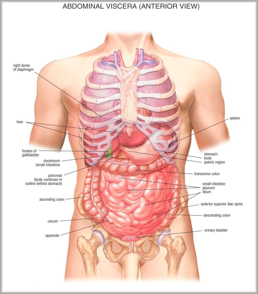
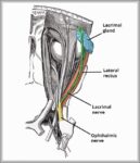
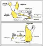
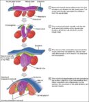
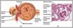
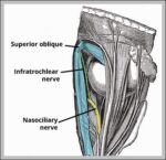
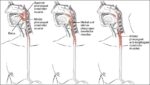
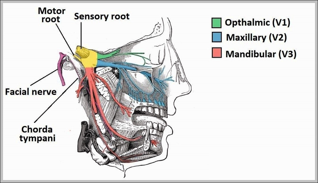
Overview of the Anatomical Distribution of the Trigeminal Nerve and its Terminal Branches Diagram
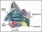
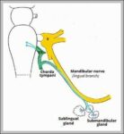
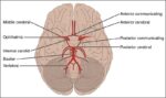
Trigeminal nerve distribution covers face via ophthalmic (V1: forehead, eye, nose), maxillary (V2: cheek, upper lip/teeth, maxillary sinus), mandibular (V3: lower jaw, lower teeth, anterior tongue, muscles of mastication). Terminal…