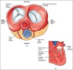
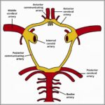
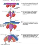
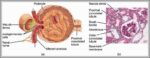
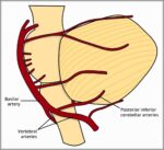
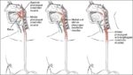
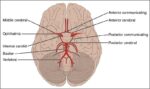
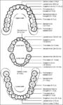
Colorectal Cancer Lg

Sometimes it is called colon cancer, for short. As the drawing shows, the colon is the large intestine or large bowel. The rectum is the passageway that connects the colon to the anus. Sometimes abnormal growths, called polyps, form in the colon or rectum. Over time, some polyps may turn into cancer.
But in some cases, changes to colon or rectal cells can cause colorectal cancer. Most often, colorectal cancer starts in gland cells that line the wall of the colon or rectum.
The colon is the final part of the digestive tract. Colon cancer typically affects older adults, though it can happen at any age. It usually begins as small, noncancerous (benign) clumps of cells called polyps that form on the inside of the colon. Over time some of these polyps can become colon cancers.

Colorectal Cancer Lg Diagram - Colorectal Cancer Lg Chart - Human anatomy diagrams and charts explained. This anatomy system diagram depicts Colorectal Cancer Lg with parts and labels. Best diagram to help learn about health, human body and medicine.

colorectal cancer lg

Colorectal Cancer Diagram

Colorectal cancer is cancer that occurs in the colon or rectum. Sometimes it is called colon cancer, for short. As the drawing shows, the colon is the large intestine or large bowel.
Pathophysiology of colorectal cancer CRC usually does not produce symptoms in early stages of the disease. If symptoms are present, they usually depend on the site of the primary tumor. Cancers of the proximal colon tend to grow larger before symptoms appear than those in the left colon and rectum.
A diagnosis of colorectal cancer either results from an assessment of a patient presenting with symptoms, or as a result of screening. The disease can be associated with spectrum of symptoms, including blood in stools, change in bowel habits and abdominal pain.

Colorectal Cancer Diagram Diagram - Colorectal Cancer Diagram Chart - Human anatomy diagrams and charts explained. This anatomy system diagram depicts Colorectal Cancer Diagram with parts and labels. Best diagram to help learn about health, human body and medicine.

colorectal cancer diagram

Colonos Screening Diagram

Colonoscopy is one of the colorectal cancer screening tests available to people in the US who are over 50 years of age.
Although widely touted in the US as the “gold standard” of colon cancer screening, colonoscopy has never been studied as a screening tool. Most of what we believe to be true about the benefits of colonoscopy have been extrapolated from randomized trials of the sigmoidoscopy.
The procedure is used as a tool for routine colon cancer screening and prevention in people beginning at age 45. Small growths called polyps can be removed during the colonoscopy before they have the chance to grow into a colorectal cancer.

Colonos Screening Diagram Diagram - Colonos Screening Diagram Chart - Human anatomy diagrams and charts explained. This anatomy system diagram depicts Colonos Screening Diagram with parts and labels. Best diagram to help learn about health, human body and medicine.

colonos screening diagram

Colon Lg Caption Figure

The figure caption labels is italic, the caption text is bold and italic. The subfigure label is bold, the text is normal and it is aligned left. As explained in the code block, singlelinecheck is turned off so that even short, one line captions use the justification setting.
Formatting captions and subcaptions in LaTeX T he captions for figures, tables, subfigures and subtables in LaTeX can be customized in various ways using the caption and subcaption packages. You can change the fonts, numbering style, alignment and format of the captions and the caption labels.
You can change the numbering or lettering style of the caption label by using variants of the following commands in your document: Each command specifies the label you want to modify (e.g. hetable) and what you want to appear as the label (e.g. alph {table}, which means to show the table counter as a lower case letter like a, b, c, etc.).

Colon Lg Caption Figure Diagram - Colon Lg Caption Figure Chart - Human anatomy diagrams and charts explained. This anatomy system diagram depicts Colon Lg Caption Figure with parts and labels. Best diagram to help learn about health, human body and medicine.

colon lg caption figure

Colon Figure

WHAT IS THE COLON? The colon is also known as the large bowel or large intestine. It is an organ that is part of the digestive system (also called the digestive tract) in the human body. The digestive system is the group of organs that allow us to eat and to use the food we …
The colon is used to express a ratio of two numbers, with no space before or after the colon.
Colorectal polyps are commonly found during standard screening exams of the colon (large intestine) and rectum (the bottom section of your colon). They affect about 20% to 30% of American adults.

Colon Figure Diagram - Colon Figure Chart - Human anatomy diagrams and charts explained. This anatomy system diagram depicts Colon Figure with parts and labels. Best diagram to help learn about health, human body and medicine.

colon figure

Colon Diagram

Colon: The long, coiled, tubelike organ that removes water from digested food. The remaining material, solid waste called stool, moves through the colon to the rectum and leaves the body through the anus. Also known as large bowel and large intestine. Image Source: MedicineNet, Inc.
The colon is the largest part of the large intestine, extending from the cecum to the rectum. It is 5 feet long and its function is to reabsorb water from digested food and concentrate solid waste material, known as stool.
The remaining material, solid waste called stool, moves through the colon to the rectum and leaves the body through the anus. Also known as large bowel and large intestine.

Colon Diagram Diagram - Colon Diagram Chart - Human anatomy diagrams and charts explained. This anatomy system diagram depicts Colon Diagram with parts and labels. Best diagram to help learn about health, human body and medicine.

colon diagram

Coloncheckmb Colon Diagram

Anatomy of Colon and Rectum. The rectum is the last anatomic segment before the anus. The ascending and descending colon are supported by peritoneal folds called mesentery. The right colon consists of the cecum, ascending colon, hepatic flexure and the right half of the transverse colon. The left colon consists of the left half…
The descending colon runs down the left side of the abdomen. The next segment, the sigmoid colon, joins the descending colon to the rectum. Stool leaves the body through an opening called the anus. Text Reference: “Anatomy of Colon and Rectum.”
Picture of Colon. The colon is made of several sections. The ascending colon travels up the right side of the abdomen. The part that sits across the abdomen is called the transverse colon. The descending colon runs down the left side of the abdomen. The next segment, the sigmoid colon, joins the descending colon to the rectum.

Coloncheckmb Colon Diagram Diagram - Coloncheckmb Colon Diagram Chart - Human anatomy diagrams and charts explained. This anatomy system diagram depicts Coloncheckmb Colon Diagram with parts and labels. Best diagram to help learn about health, human body and medicine.

coloncheckmb colon diagram

Colon Cancer Stages

There are 5 stages: stage 0 (zero) and stages I through IV (1 through 4). The stage provides a common way of describing the cancer, so doctors can work together to plan the best treatments. Here are more details on each part of the TNM system for colorectal cancer:
Stage 1: Stage 1 colon cancer indicates the cancer has grown into the inner layer of the colon, called the mucosa, to the next layer of the colon, called the submucosa. It has not spread to the lymph nodes.
The most common staging system for colorectal cancer is the TNM system. For colorectal cancer there are 5 stages – stage 0 followed by stages 1 to 4. Often the stages 1 to 4 are written as the Roman numerals I, II, III and IV.

Colon Cancer Stages Diagram - Colon Cancer Stages Chart - Human anatomy diagrams and charts explained. This anatomy system diagram depicts Colon Cancer Stages with parts and labels. Best diagram to help learn about health, human body and medicine.

colon cancer stages

Colon Cancer Screening Types Diagram

You may hear about other colorectal cancer screening tests, but Ontario’s colorectal cancer screening program, ColonCancerCheck, does not recommend using them. There is not enough research showing that they are a good way to screen for colorectal cancer. These tests include: metabolomic (blood or urine) tests.
Colorectal cancer is cancer that occurs in the colon or rectum. Sometimes it is called colon cancer, for short. As the drawing shows, the colon is the large intestine or large bowel.
Colon cancer stages, stage 4 colon cancer, stage 3 colon cancer, stage 2 colon cancer & stage 1 colon cancer. This is the earliest stage of colorectal cancer, and it is also known as cancer in situ because it is limited to the site of the tumor with no spread to adjacent structures.

Colon Cancer Screening Types Diagram Diagram - Colon Cancer Screening Types Diagram Chart - Human anatomy diagrams and charts explained. This anatomy system diagram depicts Colon Cancer Screening Types Diagram with parts and labels. Best diagram to help learn about health, human body and medicine.

colon cancer screening types diagram

Colon Cancer Screening Image

Screening is the best way of finding colorectal cancer early. Most people ages 50 to 74 are at average risk of getting colorectal cancer, meaning they do not have a first-degree relative (parent, brother, sister or child) who has been diagnosed with colorectal cancer.
Screening for Colorectal Cancer. The fecal immunochemical test (FIT) is a screening test for people at average risk of getting colorectal cancer. FIT is now used instead of the guaiac fecal occult blood test (gFOBT), which used to be Ontario’s colorectal cancer screening test.
People ages 50 to 74 without a family history of colorectal cancer who choose to be screened with flexible sigmoidoscopy should be screened every 10 years. You may hear about other colorectal cancer screening tests, but Ontario’s colorectal cancer screening program, ColonCancerCheck, does not recommend using them.

Colon Cancer Screening Image Diagram - Colon Cancer Screening Image Chart - Human anatomy diagrams and charts explained. This anatomy system diagram depicts Colon Cancer Screening Image with parts and labels. Best diagram to help learn about health, human body and medicine.

colon cancer screening image

Colon Cancer Screening

Screening is the best way of finding colorectal cancer early. Most people ages 50 to 74 are at average risk of getting colorectal cancer, meaning they do not have a first-degree relative (parent, brother, sister or child) who has been diagnosed with colorectal cancer.
If you’re between 50 and 74, you should do a home screening test every 2 years. The home screening test is the best way to prevent colorectal cancer. The home screening test can see what you cannot see – hidden blood in the stool. The home screening test can help find: colon and rectal cancer in an earlier stage making it more easily treated.
Several screening tests can be used to find polyps or colorectal cancer. The Task Force outlines the following colorectal cancer screening strategies. Talk to your doctor about which test is right for you. The guaiac-based fecal occult blood test (gFOBT) uses the chemical guaiac to detect blood in the stool.

Colon Cancer Screening Diagram - Colon Cancer Screening Chart - Human anatomy diagrams and charts explained. This anatomy system diagram depicts Colon Cancer Screening with parts and labels. Best diagram to help learn about health, human body and medicine.

colon cancer screening

Colon Cancer Explained

Colorectal cancer is a cancer that starts in the colon or the rectum. These cancers can also be named colon cancer or rectal cancer, depending on where they start. Colon cancer and rectal cancer are often grouped together because they have many features in common. Cancer starts when cells in the body start to grow out of control.
To understand colorectal cancer, it helps to know about the normal structure and function of the colon and rectum. The colon and rectum make up the large intestine (or large bowel), which is part of the digestive system, also called the gastrointestinal (GI) system (see illustration below).
As colon cancer progresses, it can grow through your colon and extend to nearby structures. The most advanced stage of colon cancer (stage IV) indicates cancer has spread to other areas of the body, such as the liver or lungs. If you’ve been diagnosed with colon cancer, your doctor may recommend tests to determine the extent (stage) of your cancer.

Colon Cancer Explained Diagram - Colon Cancer Explained Chart - Human anatomy diagrams and charts explained. This anatomy system diagram depicts Colon Cancer Explained with parts and labels. Best diagram to help learn about health, human body and medicine.

colon cancer explained

Colon Cancer Diagram

Colorectal cancer is cancer that occurs in the colon or rectum. Sometimes it is called colon cancer, for short. As the drawing shows, the colon is the large intestine or large bowel.
How is the stage determined? 1 AJCC Stage. The cancer is in its earliest stage. 2 I. The cancer has grown through the muscularis mucosa into the submucosa (T1),… 3 IIA. The cancer has grown into the outermost layers of the colon or rectum… 4 IIB. The cancer has grown through the wall of the colon or rectum but has not grown…
Pathophysiology of colorectal cancer CRC usually does not produce symptoms in early stages of the disease. If symptoms are present, they usually depend on the site of the primary tumor. Cancers of the proximal colon tend to grow larger before symptoms appear than those in the left colon and rectum.

Colon Cancer Diagram Diagram - Colon Cancer Diagram Chart - Human anatomy diagrams and charts explained. This anatomy system diagram depicts Colon Cancer Diagram with parts and labels. Best diagram to help learn about health, human body and medicine.

colon cancer diagram

Colon Cancer Colon Diagram

Colorectal cancer is cancer that occurs in the colon or rectum. Sometimes it is called colon cancer, for short. As the drawing shows, the colon is the large intestine or large bowel.
As the tumour grows in the colon or rectum, symptoms may become worse and can include: The same symptoms as above, but more severe. You may feel generally unwell, tired or lose weight. If the cancer becomes very large, it can cause a blockage (obstruction) of the colon.
Stage 1: cancer has grown through the mucosa of the colon but has not spread any further. Stage 2: Cancer has now grown into the wall of the colon. There is no lymph node involvement. Stage 3: Lymph node involvement is now seen, however, there is no metastasis to other parts of the body. Stage 4: Cancer has metastasis to other organs of the body.

Colon Cancer Colon Diagram Diagram - Colon Cancer Colon Diagram Chart - Human anatomy diagrams and charts explained. This anatomy system diagram depicts Colon Cancer Colon Diagram with parts and labels. Best diagram to help learn about health, human body and medicine.

colon cancer colon diagram

Colon Cancer

Colon cancer is sometimes called colorectal cancer, which is a term that combines colon cancer and rectal cancer, which begins in the rectum.
Most often, colorectal cancer starts in gland cells that line the wall of the colon or rectum. These gland cells make mucus that helps stool move through the colon and rectum.
A persistent change in your bowel habits, including diarrhea or constipation or a change in the consistency of your stool Many people with colon cancer experience no symptoms in the early stages of the disease. When symptoms appear, they’ll likely vary, depending on the cancer’s size and location in your large intestine.

Colon Cancer Diagram - Colon Cancer Chart - Human anatomy diagrams and charts explained. This anatomy system diagram depicts Colon Cancer with parts and labels. Best diagram to help learn about health, human body and medicine.

colon cancer

Vertebral Column Diagram

Vertebral column. The vertebral column, also known as the spinal column, is a flexible column that encloses the spinal cord and also supports the head. It consists of various groups of vertebrae and is divided into five different areas.
The spine diagram below highlights all of the vertebrae labeled. You can see the cervical vertebrae labeled at the top, the thoracic vertebrae labeled in the middle and the lumbar vertebrae labeled towards the bottom. Labeled overview of the vertebral column.
Vertebrae. The spine, vertebral column, or backbone is defined as the bony structure that runs from the inferior aspect of the occipital bone of the skull to the tip of the coccyx. However, the spinal cord is the tubular nervous tissue that travels through the vertebral canal of the vertebral column.

Vertebral Column Diagram Diagram - Vertebral Column Diagram Chart - Human anatomy diagrams and charts explained. This anatomy system diagram depicts Vertebral Column Diagram with parts and labels. Best diagram to help learn about health, human body and medicine.

vertebral column diagram

Urinary System Diagram

Urinary Tract Diagram: A gender-specific visual representation of the urinary tract, these types of diagrams can contain specific parts of the urinary tract such as: Kidney Diagram: An illustration of one or both of the kidneys.
The urinary system is composed of the kidneys, ureters, bladder and urethra. Blood from the heart travels down the aorta where it enters the kidney via the renal arteries. The kidney acts as a filter and regulator, removing waste products (urea) and balancing glucose, electrolytes (salt, potassium and other minerals) and water levels in the blood.
How does the urinary system work? The urinary system’s function is to filter blood and create urine as a waste by-product. The organs of the urinary system include the kidneys, renal pelvis, ureters, bladder and urethra. The body takes nutrients from food and converts them to energy.

Urinary System Diagram Diagram - Urinary System Diagram Chart - Human anatomy diagrams and charts explained. This anatomy system diagram depicts Urinary System Diagram with parts and labels. Best diagram to help learn about health, human body and medicine.

urinary system diagram

Lymph Capillary Diagram

Structure of Lymphatic Capillaries. Lymph or lymphatic capillaries are tiny thin-walled vessels, closed at one end and located in the spaces between cells throughout the body. These are particularly dense within connective tissue. Lymphatic capillaries are slightly larger in diameter than blood capillaries and contain flap-like “minivalves”…
Upon entering the lumen of a lymphatic capillary, the collected fluid and associated cells (notably white blood cells) is known as lymph. Each lymphatic capillary carries lymph into a lymphatic vessel, which in turn connects to a lymph node. Lymph is ultimately returned to the venous circulation .
Because lymphatic capillaries have a closed end and minivalves normally prevent backflow into tissues, the pressure of lymph becomes higher as more lymph is collected from the tissues, which sends the lymph fluid forward. Multiple capillaries converge in collecting vessels, where the internal valves and smooth muscle start to appear.

Lymph Capillary Diagram Diagram - Lymph Capillary Diagram Chart - Human anatomy diagrams and charts explained. This anatomy system diagram depicts Lymph Capillary Diagram with parts and labels. Best diagram to help learn about health, human body and medicine.

lymph capillary diagram

Lower Extremity Muscles Diagram

The primary muscle in this part of the body is the gastrocnemius, which gives the calf its signature bulging, muscular appearance. The anterior tibial, posterior tibial and fibular arteries are responsible for blood supply to the lower leg. The lower leg makes up a large portion of an individual’s overall body weight.
Key facts about the lower extremity Hip and pelvis Bones: hip bones, saccrum, coccyx Hip jo … Thigh Bones: femur Joints: hip and knee Muscle … Knee Bones: tibia, fibula, patella Type: hing … Leg Bones: tibia, fibula Joints: knee and an … Ankle and foot Ankle joint: hinged joint capable of …
The leg muscles and tendons produce tension, stabilize the joints of the legs, and create movement. The main muscle groups in the legs are: quadriceps, hamstrings, adductors in the upper leg or thigh, and the calves in the lower legs.

Lower Extremity Muscles Diagram Diagram - Lower Extremity Muscles Diagram Chart - Human anatomy diagrams and charts explained. This anatomy system diagram depicts Lower Extremity Muscles Diagram with parts and labels. Best diagram to help learn about health, human body and medicine.

lower extremity muscles diagram

Intestine Diagram

The specific anatomy of the large intestines is best described by referencing the individual parts, which are described in more detail below. The large intestine is roughly a meter and a half (about four and a half feet) in total length from the end of the small intestine to the anus.
8,204 intestine diagram stock photos, vectors, and illustrations are available royalty-free.
The large intestine anatomy may appear segmented due to bumps called haustra. The large intestine’s main function is to absorb any remaining nutrients and water in the chyme received from the small intestine. It also forms stool and transports waste out of the body.

Intestine Diagram Diagram - Intestine Diagram Chart - Human anatomy diagrams and charts explained. This anatomy system diagram depicts Intestine Diagram with parts and labels. Best diagram to help learn about health, human body and medicine.

intestine diagram