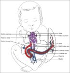
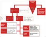
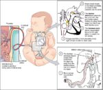
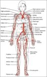
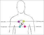
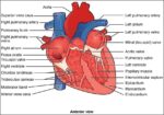
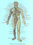
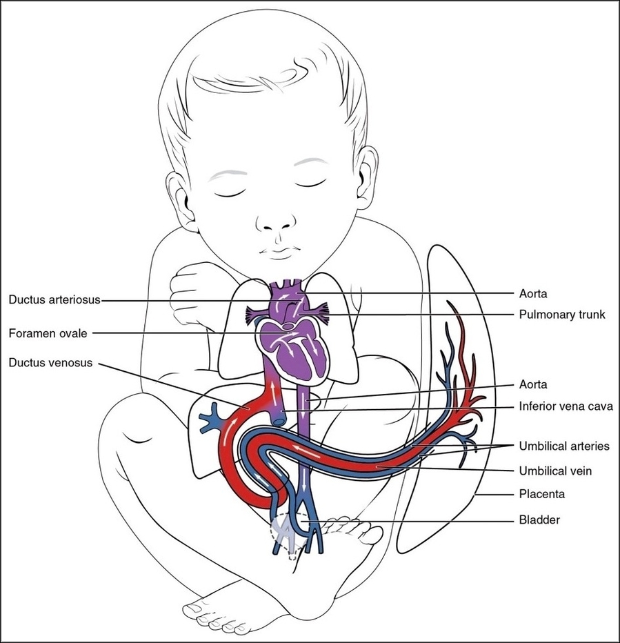
Cardiovascular System Blood Vessels Image

Blood Vessels. Blood vessels are the body’s highways that allow blood to flow quickly and efficiently from the heart to every region of the body and back again. The size of blood vessels corresponds with the amount of blood that passes through the vessel.
The blood circulatory system (cardiovascular system) delivers nutrients and oxygen to all cells in the body. It consists of the heart and the blood vessels running through the entire body. NCBI Skip to main content Skip to navigation Resources How To About NCBI Accesskeys

Cardiovascular System Blood Vessels Image Diagram - Cardiovascular System Blood Vessels Image Chart - Human anatomy diagrams and charts explained. This anatomy system diagram depicts Cardiovascular System Blood Vessels Image with parts and labels. Best diagram to help learn about health, human body and medicine.

Cardiovascular System Blood Vessels Image

Pic Of Human Anatomy Image

28,679 anatomy of the human body stock photos and images available, or start a new search to explore more stock photos and images.
Abstract image human body in the form of a starry sky or space, consisting of points, lines, and shapes in the form of planets, stars and the universe. Low poly vector background. human body anatomy stock illustrations Abstract image human body in the form of a starry sky or space,…

Pic Of Human Anatomy Image Diagram - Pic Of Human Anatomy Image Chart - Human anatomy diagrams and charts explained. This anatomy system diagram depicts Pic Of Human Anatomy Image with parts and labels. Best diagram to help learn about health, human body and medicine.

Pic Of Human Anatomy Image

Human Anatomy Torso Image

9,591 human torso anatomy stock photos and images available, or search for human anatomy to find more great stock photos and pictures.
This resource is aimed at helping anyone within the medical community with illustrations for articles, blogs, assignments, and posters. The high-quality vector images of the human anatomy are available to everyone who wants to use them, free of charge.

Human Anatomy Torso Image Diagram - Human Anatomy Torso Image Chart - Human anatomy diagrams and charts explained. This anatomy system diagram depicts Human Anatomy Torso Image with parts and labels. Best diagram to help learn about health, human body and medicine.

Human Anatomy Torso Image

Muscle Cell Diagram Image

A muscle cell is a long cell compared to other forms of cells, and many muscle cells connect together to form the long fibers found in muscle tissue. As seen in the image below, a muscle cell is a compact bundle of many myofibrils.
1,416 muscle cell stock photos and images available, or search for smooth muscle cell or skeletal muscle cell to find more great stock photos and pictures. Sem Of Muscle Fiber.

Muscle Cell Diagram Image Diagram - Muscle Cell Diagram Image Chart - Human anatomy diagrams and charts explained. This anatomy system diagram depicts Muscle Cell Diagram Image with parts and labels. Best diagram to help learn about health, human body and medicine.

Muscle Cell Diagram Image

Images Of Anatomy Image

132,206 anatomy stock photos and images available or search for anatomy model or muscle anatomy to find more great stock photos and pictures.
Human Anatomy. Is primarily the scientific study of the morphology of the human body. Anatomy is subdivided into gross anatomy and microscopic anatomy &#x28 Watercolor anatomy organ heart illustration. High quality illustration human body organs heart for education Dog Nervous System – Canis Lupus Familiaris Anatomy – isolated o. N white

Images Of Anatomy Image Diagram - Images Of Anatomy Image Chart - Human anatomy diagrams and charts explained. This anatomy system diagram depicts Images Of Anatomy Image with parts and labels. Best diagram to help learn about health, human body and medicine.

Images Of Anatomy Image scaled

All Body Muscles Image

94,749 muscles body stock photos and images available, or search for oxygen or the human body to find more great stock photos and pictures.
Forearms – Anatomy Muscles Rhomboid minor and rhomboid major, levator scapulae and latissimus dorsi muscles – didactic board of anatomy of human bony and muscular system, posterior view Abs – Anatomy Muscles Leg muscles of the man Human Body Organs (Lungs) 3D Triceps – Anatomy Muscles Hand muscle connection with brain

All Body Muscles Image Diagram - All Body Muscles Image Chart - Human anatomy diagrams and charts explained. This anatomy system diagram depicts All Body Muscles Image with parts and labels. Best diagram to help learn about health, human body and medicine.

All Body Muscles Image

Human Body Back View Image

52,129 human back anatomy stock photos, vectors, and illustrations are available royalty-free.
Human anatomy xray view of intestines, on light back. Human anatomy xray view of intestines, showing stomach, colon, intestines, lungs, urinary system and Andrenal. The internal andrenal Organs – back view

Human Body Back View Image Diagram - Human Body Back View Image Chart - Human anatomy diagrams and charts explained. This anatomy system diagram depicts Human Body Back View Image with parts and labels. Best diagram to help learn about health, human body and medicine.

Human Body Back View Image

Male Body Parts Image

2,101,867 male body parts stock photos and images available, or start a new search to explore more stock photos and images. Male External Genital Organs. See Images 0348407 For The Male External Genital Organs, 0348607 And 0348707 For The Female External Genital Organs.
What are the external parts? Your penis and scrotum are the two parts of the male (or what’s typically called male) external sex anatomy (outside your body). Your penis is made of 3 layers of spongy tissue. When you get turned on, blood fills these tissues.

Male Body Parts Image Diagram - Male Body Parts Image Chart - Human anatomy diagrams and charts explained. This anatomy system diagram depicts Male Body Parts Image with parts and labels. Best diagram to help learn about health, human body and medicine.

Male Body Parts Image

Muscular System Parts Image

This muscular system picture shows all the major muscle groups on the human body from the frontal view. To see a muscular system diagram from the posterior (back) view click here.
Muscular System Parts. 1 Skeletal Muscle. Striated muscle, or Skeletal muscle, is the tissue most commonly associated with the muscular system. This type of muscle attaches to … 2 Visceral Muscle. 3 Cardiac Muscle.

Muscular System Parts Image Diagram - Muscular System Parts Image Chart - Human anatomy diagrams and charts explained. This anatomy system diagram depicts Muscular System Parts Image with parts and labels. Best diagram to help learn about health, human body and medicine.

Muscular System Parts Image

Anatomy Of The Wrist And Hand Image

Anatomy of the Hand and Wrist: Bones, Muscles, Tendons, Nerves. The wrist links the hand to the arm. The wrist is a complex mechanical system of 8 small bones known as the carpal bones. The carpal bones are arranged in 2 interrelated rows. One row connects with the ends of the bones in the forearm— radius and ulna.
Wrist anatomy is the study of the bones, ligaments and other structures in the wrist. The wrist joint is a complex joint which connects the forearm to the hand, allowing a wide range of movement. However, it is susceptible to injury, especially from repetitive strain.

Anatomy Of The Wrist And Hand Image Diagram - Anatomy Of The Wrist And Hand Image Chart - Human anatomy diagrams and charts explained. This anatomy system diagram depicts Anatomy Of The Wrist And Hand Image with parts and labels. Best diagram to help learn about health, human body and medicine.

Anatomy Of The Wrist And Hand Image

Lvn Nurse Image

As an LVN nurse you can get paid for taking care of people while getting the valuable experience you need to become an RN. What Is an LVN or LPN? Job Description & Career Guide Learn how to become an LVN (licensed vocational nurse) in this comprehensive guide. It covers what is an LVN, the required education, average salaries, and its job outlook.
A Licensed Practical Nurse (LPN), or Licensed Vocational Nurse (LVN), is an entry-level nurse with a practical education. To become an LPN, you must complete high school, finish 1 year of full-time nursing education, and pass the NCLEX exam.

Lvn Nurse Image Diagram - Lvn Nurse Image Chart - Human anatomy diagrams and charts explained. This anatomy system diagram depicts Lvn Nurse Image with parts and labels. Best diagram to help learn about health, human body and medicine.

Lvn Nurse Image

Body Organ Diagram Woman Image

465 female anatomy diagram stock photos and images available, or start a new search to explore more stock photos and images. Figures illustrating the difference between a natural waist and a constricted waist deformed through the use of corsets, from a published essay by…
Woman anatomy cardiovascular system with skeleton and internal organs, rear and front views. An illustration depicting the major organs in the human body, as well as a ghosted view of the bones of the skeleton. Full length view of male human body with organs.

Body Organ Diagram Woman Image Diagram - Body Organ Diagram Woman Image Chart - Human anatomy diagrams and charts explained. This anatomy system diagram depicts Body Organ Diagram Woman Image with parts and labels. Best diagram to help learn about health, human body and medicine.

Body Organ Diagram Woman Image

System Of The Body Image

Abstract image of a human body in the form of a starry sky or space, consisting of points, lines, and shapes in the form of planets, stars and the universe. Low poly vector background. the human body stock illustrations
183,298 human body systems stock photos, vectors, and illustrations are available royalty-free.

System Of The Body Image Diagram - System Of The Body Image Chart - Human anatomy diagrams and charts explained. This anatomy system diagram depicts System Of The Body Image with parts and labels. Best diagram to help learn about health, human body and medicine.

System Of The Body Image

Coccyx Bone Image

The coccyx is a triangular arrangement of bone that makes up the very bottom portion of the spine below the sacrum. It represents a vestigial tail, hence the common term tailbone. See Normal Spinal Anatomy
Diagnosis of coccyx pain. The steps to diagnose the exact cause of the coccyx pain are as follows: (a)Physical examination: Inspection of the tailbone area for any visible bruises or lumps. External palpation to check for tailbone tenderness or crackling sound. Internal palpation is done through the rectum.

Coccyx Bone Image Diagram - Coccyx Bone Image Chart - Human anatomy diagrams and charts explained. This anatomy system diagram depicts Coccyx Bone Image with parts and labels. Best diagram to help learn about health, human body and medicine.

Coccyx Bone Image

Uterus Anatomy Pictures Image

Uterus Anatomy Pictures Image Diagram - Uterus Anatomy Pictures Image Chart - Human anatomy diagrams and charts explained. This anatomy system diagram depicts Uterus Anatomy Pictures Image with parts and labels. Best diagram to help learn about health, human body and medicine.

Uterus Anatomy Pictures Image

Sperm Cells Image

Sperm cells are gametes (sex cells) that are produced in the testicular organ (gonad) of male human beings and animals. Like the female gamete (oocyte), sperm cells carry a total of 23 chromosomes that are a result of a process known as meiosis. In both animals and human beings, among many other organisms, these cells are involved in …
4,744 sperm cell stock photos and images available, or start a new search to explore more stock photos and images. Human spermatozoa.

Sperm Cells Image Diagram - Sperm Cells Image Chart - Human anatomy diagrams and charts explained. This anatomy system diagram depicts Sperm Cells Image with parts and labels. Best diagram to help learn about health, human body and medicine.

Sperm Cells Image

Picture Of Human Hip Bones Image

1,047 hip bone stock photos and images available, or search for hip bone icon or hip bone 3d to find more great stock photos and pictures.
The hip is formed where the thigh bone (femur) meets the three bones that make up the pelvis: the ilium, the pubis (pubic bone) and the ischium. These three bones converge to form the acetabulum, a deep socket on the outer edge of the pelvis. By adulthood, these three bones are completely fused and the pelvis is effectively a single bone.

Picture Of Human Hip Bones Image Diagram - Picture Of Human Hip Bones Image Chart - Human anatomy diagrams and charts explained. This anatomy system diagram depicts Picture Of Human Hip Bones Image with parts and labels. Best diagram to help learn about health, human body and medicine.

Picture Of Human Hip Bones Image

Function Of The Thyroid Gland Image

Thyroid gland. Medically reviewed by Healthline’s Medical Network on February 18, 2015. The thyroid gland covers the windpipe from three sides. Two hormones of the thyroid gland, T4 (thyroxine) and T3 (triiodothyronine), help the body to produce and regulate the hormones adrenaline (also called epinephrine) and dopamine.
The thyroid releases them into your bloodstream, which takes them through your body. Another gland, called the pituitary gland, tells your thyroid how much of these hormones your body needs. Sometimes, the thyroid gland develops a problem.

Function Of The Thyroid Gland Image Diagram - Function Of The Thyroid Gland Image Chart - Human anatomy diagrams and charts explained. This anatomy system diagram depicts Function Of The Thyroid Gland Image with parts and labels. Best diagram to help learn about health, human body and medicine.

Function Of The Thyroid Gland Image

Adrenal Gland Diagram Image

Adrenal glands sit atop the kidneys and are composed of an outer cortex and an inner medulla, which produce different types of hormones. Human endocrine system adrenal gland stock illustrations
5,601 adrenal gland stock photos, vectors, and illustrations are available royalty-free.

Adrenal Gland Diagram Image Diagram - Adrenal Gland Diagram Image Chart - Human anatomy diagrams and charts explained. This anatomy system diagram depicts Adrenal Gland Diagram Image with parts and labels. Best diagram to help learn about health, human body and medicine.

Adrenal Gland Diagram Image

Human Knee Anatomy Diagram Image

3,344 knee anatomy stock photos and images available, or search for knee anatomy illustration or human knee anatomy to find more great stock photos and pictures.
Knee joint anatomy involves looking at each of the different structures in and around the knee. The knee joint is the largest and one of the most complex joints in the human body.

Human Knee Anatomy Diagram Image Diagram - Human Knee Anatomy Diagram Image Chart - Human anatomy diagrams and charts explained. This anatomy system diagram depicts Human Knee Anatomy Diagram Image with parts and labels. Best diagram to help learn about health, human body and medicine.

Human Knee Anatomy Diagram Image