Picture Of A Human Torso Image

9,591 human torso anatomy stock photos and images available, or search for human anatomy to find more great stock photos and pictures.
Male Torso stock illustrations A torso of human body in charcoal style. Include AI,PDF and CDR files. Still Life-Mannequin A Two Mannequins photographed in a still life setting against a grey wall.

Picture Of A Human Torso Image Diagram - Picture Of A Human Torso Image Chart - Human anatomy diagrams and charts explained. This anatomy system diagram depicts Picture Of A Human Torso Image with parts and labels. Best diagram to help learn about health, human body and medicine.

Picture Of A Human Torso Image

Human Body Stomach Image

2,755 human stomach anatomy stock photos and images available or start a new search to explore more stock photos and images.
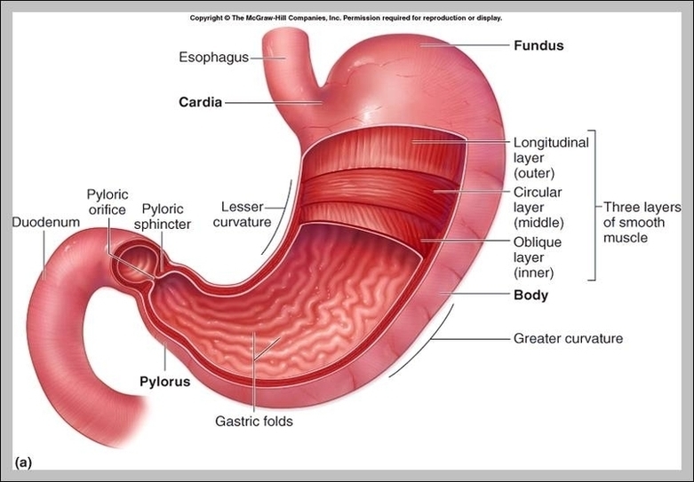
Next. The stomach is a muscular organ located on the left side of the upper abdomen. The stomach receives food from the esophagus. As food reaches the end of the esophagus, it enters the stomach …

Human Body Stomach Image Diagram - Human Body Stomach Image Chart - Human anatomy diagrams and charts explained. This anatomy system diagram depicts Human Body Stomach Image with parts and labels. Best diagram to help learn about health, human body and medicine.

Human Body Stomach Image

Study Anatomy Online Image

Common ways to learn anatomy online include YouTube videos and online multimedia learning platforms such as Kenhub. There are several fantastic YouTube channels available for learning anatomy. For those who don’t enjoy the traditional textbook approach to learning, they’re a great alternative.
Everything you need to know about how to study anatomy. Now all that’s left to do is actually study! Learning anatomy is not an easy task, but with the right tools and the right mindset, you’ll get there. We hope this article has given you some inspiration on how and where to get started.

Study Anatomy Online Image Diagram - Study Anatomy Online Image Chart - Human anatomy diagrams and charts explained. This anatomy system diagram depicts Study Anatomy Online Image with parts and labels. Best diagram to help learn about health, human body and medicine.

Study Anatomy Online Image

Image Of Human Body Image

Abstract image human body in the form of a starry sky or space, consisting of points, lines, and shapes in the form of planets, stars and the universe. Low poly vector background. human body anatomy stock illustrations Abstract image human body in the form of a starry sky or space,…
130,635 inside the human body stock photos and images available, or search for human anatomy or blood cells to find more great stock photos and pictures.

Image Of Human Body Image Diagram - Image Of Human Body Image Chart - Human anatomy diagrams and charts explained. This anatomy system diagram depicts Image Of Human Body Image with parts and labels. Best diagram to help learn about health, human body and medicine.

Image Of Human Body Image

Hinge Joints In The Body Image

A hinge joint is a common class of synovial joint that includes the ankle, elbow, and knee joints. Hinge joints are formed between two or more bones where the bones can only move along one axis to flex or extend.
The elbows, the knees, and the middle and end joints of the fingers are hinge joints. Hinge joints are prone to injury when lateral forces are applied to the joint. Many knee injuries occur in this manner, according to the National Institute of Arthritis and Musculoskeletal and Skin Diseases.

Hinge Joints In The Body Image Diagram - Hinge Joints In The Body Image Chart - Human anatomy diagrams and charts explained. This anatomy system diagram depicts Hinge Joints In The Body Image with parts and labels. Best diagram to help learn about health, human body and medicine.

Hinge Joints In The Body Image

Skeletal Pictures Image

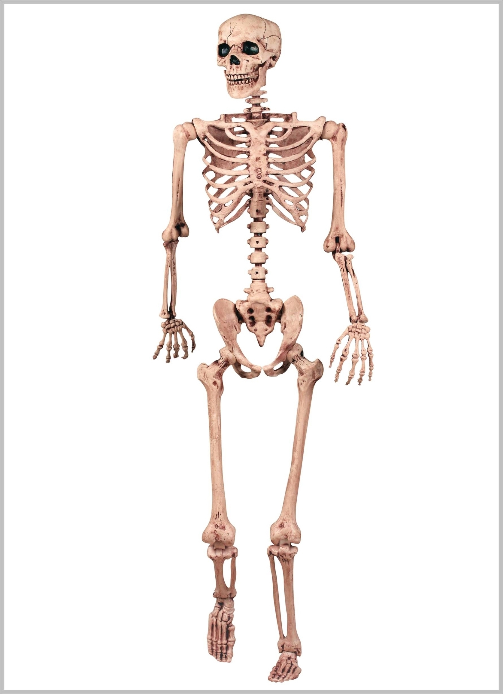
45,936 human skeleton stock photos and images available, or search for human skeleton anatomy or human skeleton vector to find more great stock photos and pictures.
Skeleton in various poses is cartoon style Skeleton in various poses is cartoon style. Funny skeleton with various gestures. Vector illustration. human skeleton stock illustrations Skeleton in various poses is cartoon style.

Skeletal Pictures Image Diagram - Skeletal Pictures Image Chart - Human anatomy diagrams and charts explained. This anatomy system diagram depicts Skeletal Pictures Image with parts and labels. Best diagram to help learn about health, human body and medicine.

Skeletal Pictures Image

Taste Buds On Tongue Diagram Image

Taste bud and the papillae of the tongue. basic taste areas: sweet, salty, sour, bitter and umami. Taste bud and the papillae of the tongue. Human mouth isolated Taste bud. Mature and Immature taste Receptor, Support and Basal Cells, Epithelium Of tongue.
Taste buds are a small organ located primarily on the tongue. The adult human tongue contains between 2,000 and 8,000 taste buds, each of which are made up of 50 to 150 taste receptor cells. Taste receptor cells are responsible for reporting the sense of taste to the brain .

Taste Buds On Tongue Diagram Image Diagram - Taste Buds On Tongue Diagram Image Chart - Human anatomy diagrams and charts explained. This anatomy system diagram depicts Taste Buds On Tongue Diagram Image with parts and labels. Best diagram to help learn about health, human body and medicine.

Taste Buds On Tongue Diagram Image

Muscles In Forearm Image

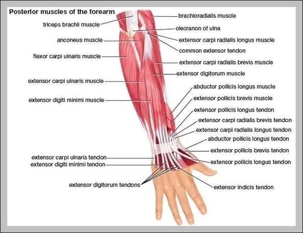
The muscles of the forearm can be divided into two groups: anterior (flexors) and posterior (extensors). Both the flexors and extensors are further divided into superficial and deep layers. The forearm muscles that control the movement of the hands are known as extrinsic hand muscles.
27,539 arm muscle anatomy stock photos, vectors, and illustrations are available royalty-free.

Muscles In Forearm Image Diagram - Muscles In Forearm Image Chart - Human anatomy diagrams and charts explained. This anatomy system diagram depicts Muscles In Forearm Image with parts and labels. Best diagram to help learn about health, human body and medicine.

Muscles In Forearm Image

Labelled Diagram Of The Eye Image

Use your mouse or finger to hover over a box to highlight the part to be named. Drag and drop the text labels onto the boxes next to the eye diagram If you want to redo an answer, click on the box and the answer will go back to the top so you can move it to another box.
The Human Eye (Eyeball) Diagram, Parts and Pictures. The human eye consists of the eyeball, optic nerve, orbit and appendages (eyelids, extraocular muscles and lacrimal glands). While the eyeball is the actual sensory organ, the other parts of of the eye are equally important in maintaining the health and function of the eye as a whole.

Labelled Diagram Of The Eye Image Diagram - Labelled Diagram Of The Eye Image Chart - Human anatomy diagrams and charts explained. This anatomy system diagram depicts Labelled Diagram Of The Eye Image with parts and labels. Best diagram to help learn about health, human body and medicine.

Labelled Diagram Of The Eye Image 1

Muscles In The Foot Image

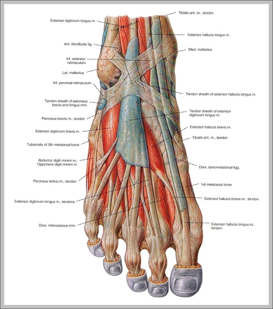
The muscles of the foot can be split into two groups, the extrinsic and intrinsic muscles. The extrinsic foot muscles are found in the lower leg and act to dorsiflex, plantarflex, invert and evert the foot. The intrinsic foot muscles are entirely contained within it, and primarily act to move the toes.
The 20-plus muscles in the foot help enable movement, while also giving the foot its shape. Like the fingers, the toes have flexor and extensor muscles that power their movement and play a large role in balance. However, many muscles that power the foot’s movement originate as high up as the back of the knee.

Muscles In The Foot Image Diagram - Muscles In The Foot Image Chart - Human anatomy diagrams and charts explained. This anatomy system diagram depicts Muscles In The Foot Image with parts and labels. Best diagram to help learn about health, human body and medicine.

Muscles In The Foot Image

Human Vocal Organ Anatomy

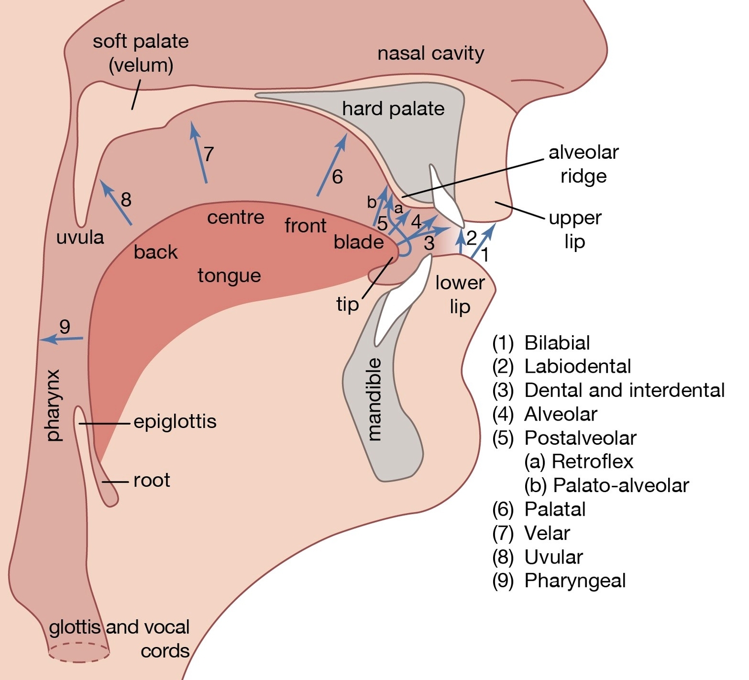
This lesson goes over the anatomy of the vocal tract, the amazing part of our body responsible for producing human speech. We will cover the larynx and the pharynx, the major components of our vocal tract. This morning as you were singing in the shower (it’s okay, no one heard), did you stop to think about how that sound was produced?
The vocal cords are located just above the trachea, below the epiglottis. The difference in the size of vocal cords of the males and females is what gives rise to different pitches of voices. In females, the vocal cords can measure anywhere in between 12 and 17 mm while in males it can vary from 17 to 25 mm.

Human Vocal Organ Anatomy Diagram - Human Vocal Organ Anatomy Chart - Human anatomy diagrams and charts explained. This anatomy system diagram depicts Human Vocal Organ Anatomy with parts and labels. Best diagram to help learn about health, human body and medicine.

human vocal organ anatomy

How To Become A Healthcare Executive Image

This popular certification for hospital executives requires: Holding a current position, as well as five years of experience in healthcare management Obtaining two references, including an interview with a current fellow and a written reference from a colleague
In some circumstances, healthcare professionals can become hospital CEOs. Given that healthcare experience is one requirement for health leadership roles, some doctors and nurses have accelerated paths to hospital CEO roles.

How To Become A Healthcare Executive Image Diagram - How To Become A Healthcare Executive Image Chart - Human anatomy diagrams and charts explained. This anatomy system diagram depicts How To Become A Healthcare Executive Image with parts and labels. Best diagram to help learn about health, human body and medicine.

How To Become A Healthcare Executive Image

Human Skeleton With Bones Labeled Image

31,815 human skeleton anatomy stock photos and images available, or search for human bones or human anatomy to find more great stock photos and pictures.
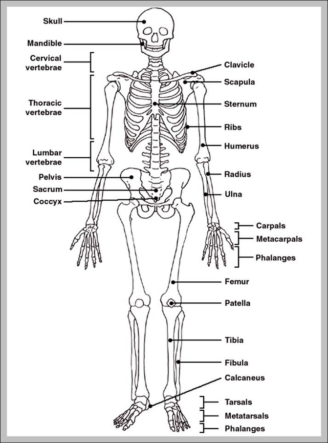
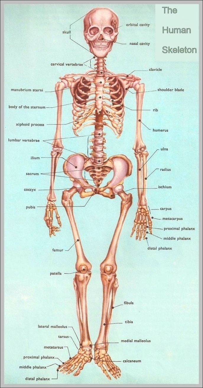
62 labeled diagram of the skeletal system pic stock photos and images available, or start a new search to explore more stock photos and images. Skeletal Poster Human skeletal system poster containing detailed information about the skeletal structure.

Human Skeleton With Bones Labeled Image Diagram - Human Skeleton With Bones Labeled Image Chart - Human anatomy diagrams and charts explained. This anatomy system diagram depicts Human Skeleton With Bones Labeled Image with parts and labels. Best diagram to help learn about health, human body and medicine.

Human Skeleton With Bones Labeled Image

Anatomy Human Arm Image

4,995 human arm anatomy stock photos and images available, or start a new search to explore more stock photos and images.
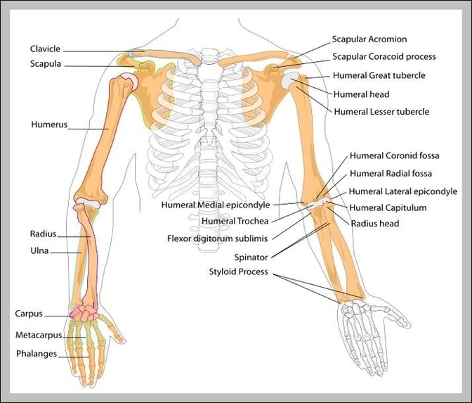
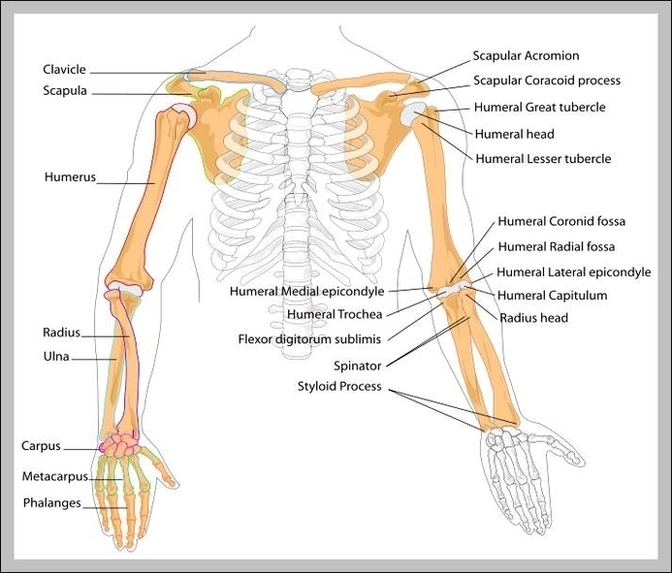
The human arm is divided into two main regions, the portion from the elbow to the wrist known as the forearm, and the segment from the shoulder to the elbow referred to as the arm. Arm muscle anatomy enables the arm to perform a variety of movements, including flexion, extension, pronation, and supination.

Anatomy Human Arm Image Diagram - Anatomy Human Arm Image Chart - Human anatomy diagrams and charts explained. This anatomy system diagram depicts Anatomy Human Arm Image with parts and labels. Best diagram to help learn about health, human body and medicine.
Anatomy Human Arm Image

Picture Of Human Image

Abstract image human body in the form of a starry sky or space, consisting of points, lines, and shapes in the form of planets, stars and the universe. Low poly vector background. human body anatomy stock illustrations Abstract image human body in the form of a starry sky or space,…
28,679 anatomy of the human body stock photos and images available, or start a new search to explore more stock photos and images.

Picture Of Human Image Diagram - Picture Of Human Image Chart - Human anatomy diagrams and charts explained. This anatomy system diagram depicts Picture Of Human Image with parts and labels. Best diagram to help learn about health, human body and medicine.

Picture Of Human Image

Sternum Xiphoid Process Image

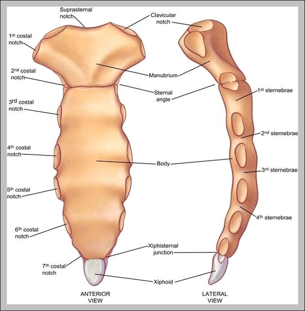
Sternum Xiphoid Process Image Diagram - Sternum Xiphoid Process Image Chart - Human anatomy diagrams and charts explained. This anatomy system diagram depicts Sternum Xiphoid Process Image with parts and labels. Best diagram to help learn about health, human body and medicine.

Sternum Xiphoid Process Image

Diagram Of The Muscle System Image

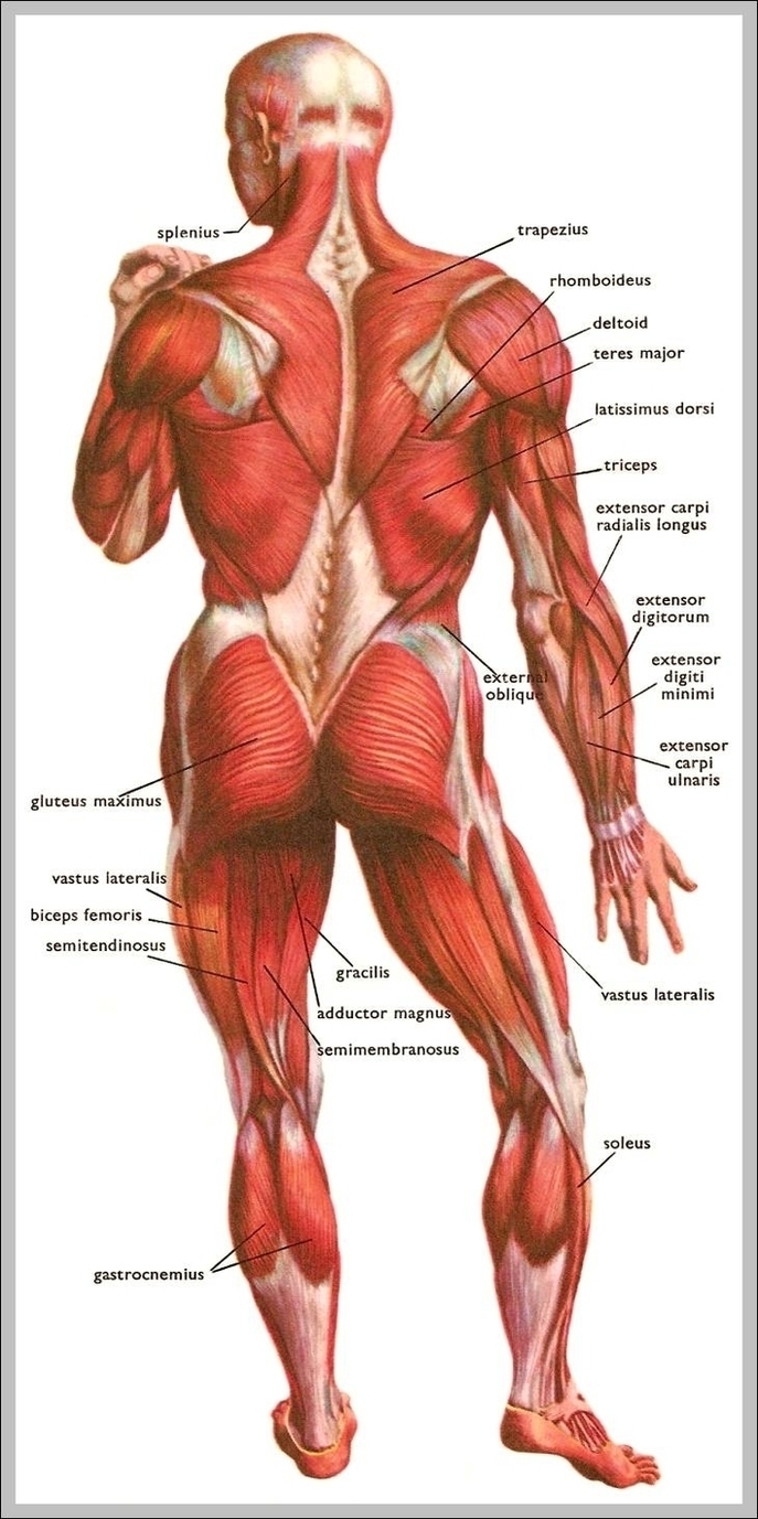
101,842 muscular system stock photos and images available, or search for muscle anatomy or abstract pattern background to find more great stock photos and pictures.
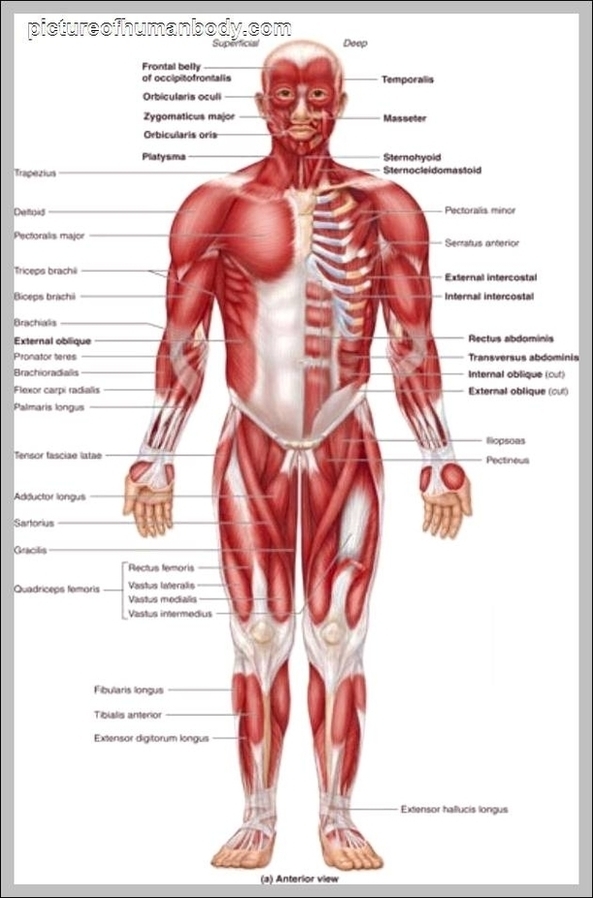
Last Updated: Jul 16, 2019 The muscular system is responsible for the movement of the human body. Attached to the bones of the skeletal system are about 700 named muscles that make up roughly half of a person’s body weight. Each of these muscles is a discrete organ constructed of skeletal muscle tissue, blood vessels, tendons, and nerves.

Diagram Of The Muscle System Image Diagram - Diagram Of The Muscle System Image Chart - Human anatomy diagrams and charts explained. This anatomy system diagram depicts Diagram Of The Muscle System Image with parts and labels. Best diagram to help learn about health, human body and medicine.

Diagram Of The Muscle System Image

Human Eye Diagram For Kids Image

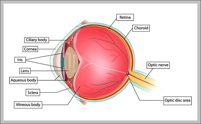
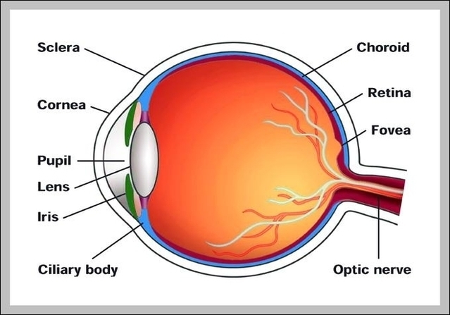
Dimensions:760 x 772 Photo description: This human eye diagram gives an excellent overview of the human eye. The cross section features labeled parts such as the iris, pupil, cornea, lens, retina, choroid, optic disc, optic nerve and fovea.
Human Eye Diagram: Contrary to popular belief, the eyes are not perfectly spherical; instead, it is made up of two separate segments fused together. Explore: Facts About The Eye To understand more in detail about our eye and how our eye functions, we need to look into the structure of the human eye.

Human Eye Diagram For Kids Image Diagram - Human Eye Diagram For Kids Image Chart - Human anatomy diagrams and charts explained. This anatomy system diagram depicts Human Eye Diagram For Kids Image with parts and labels. Best diagram to help learn about health, human body and medicine.

Human Eye Diagram For Kids Image

Diagram Of Human Arm Image

The human arm is divided into two main regions, the portion from the elbow to the wrist known as the forearm, and the segment from the shoulder to the elbow referred to as the arm. Arm muscle anatomy enables the arm to perform a variety of movements, including flexion, extension, pronation, and supination.
Each of these two sections of the human arm consists of arm muscle anatomy that allows for the flexion, extension, pronation, and supination of the arm, as will be further discussed below. While four muscles are responsible for the upper arm musculature, there are over twenty muscles that move the forearm, wrist, hands, and fingers.

Diagram Of Human Arm Image Diagram - Diagram Of Human Arm Image Chart - Human anatomy diagrams and charts explained. This anatomy system diagram depicts Diagram Of Human Arm Image with parts and labels. Best diagram to help learn about health, human body and medicine.

Diagram Of Human Arm Image

Labeled Body Parts In Spanish Image

The names of body parts are used much the same as they are in Spanish as in English, but with one significant difference. In Spanish, names of parts of the body are frequently preceded by the definite article (el, la, los or las, meaning “the”) instead of possessive adjectives (such as mi for “my” and tu for “your”).
I broke my [body part]. (Ex. I broke my arm.) Me rompí [body part]. (Ex. Me rompí el brazo.) I cut my finger. Me corto el dedo. I have a pain/ache of [body part]. (Ex. I have a headache.)** Tengo dolor de [body part]. (Ex.Tengo dolor de cabeza.) I have black/blond/brown/red/gray hair.

Labeled Body Parts In Spanish Image Diagram - Labeled Body Parts In Spanish Image Chart - Human anatomy diagrams and charts explained. This anatomy system diagram depicts Labeled Body Parts In Spanish Image with parts and labels. Best diagram to help learn about health, human body and medicine.
Labeled Body Parts In Spanish Image