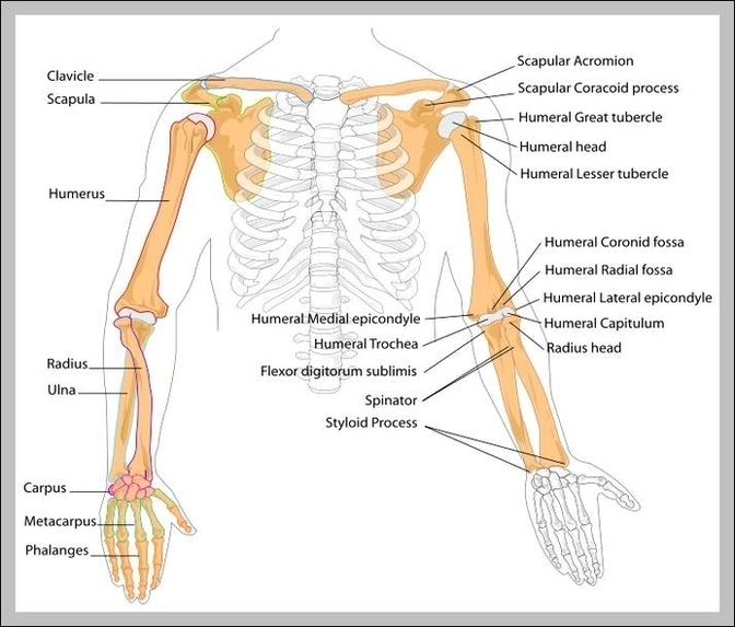
Diagram Of Human Arm Image

The human arm is divided into two main regions, the portion from the elbow to the wrist known as the forearm, and the segment from the shoulder to the elbow referred to as the arm. Arm muscle anatomy enables the arm to perform a variety of movements, including flexion, extension, pronation, and supination.
Each of these two sections of the human arm consists of arm muscle anatomy that allows for the flexion, extension, pronation, and supination of the arm, as will be further discussed below. While four muscles are responsible for the upper arm musculature, there are over twenty muscles that move the forearm, wrist, hands, and fingers.
Each of these two sections of the human arm consists of arm muscle anatomy that allows for the flexion, extension, pronation, and supination of the arm, as will be further discussed below. While four muscles are responsible for the upper arm musculature, there are over twenty muscles that move the forearm, wrist, hands, and fingers.
