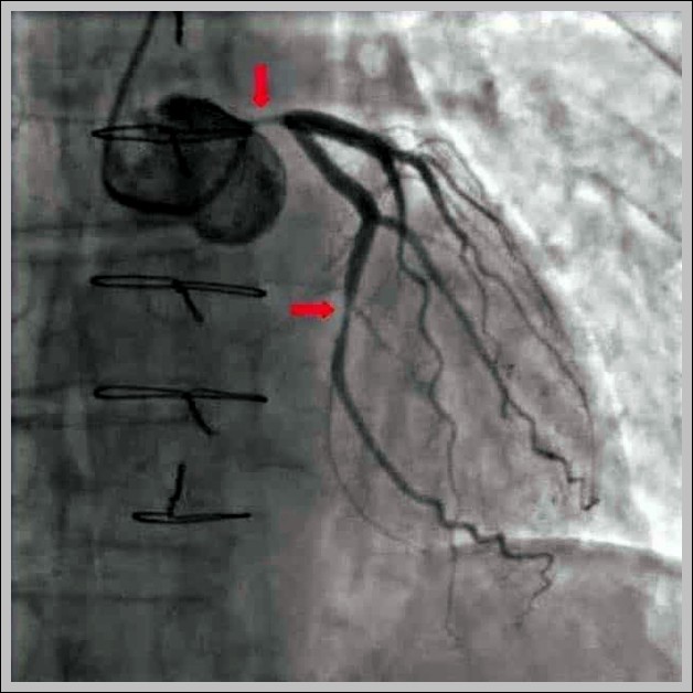
Coronary angiograms label narrowing (stenosis) in arteries like LAD, circumflex, right coronary, often graded by percent occlusion to guide stenting or bypass decisions.
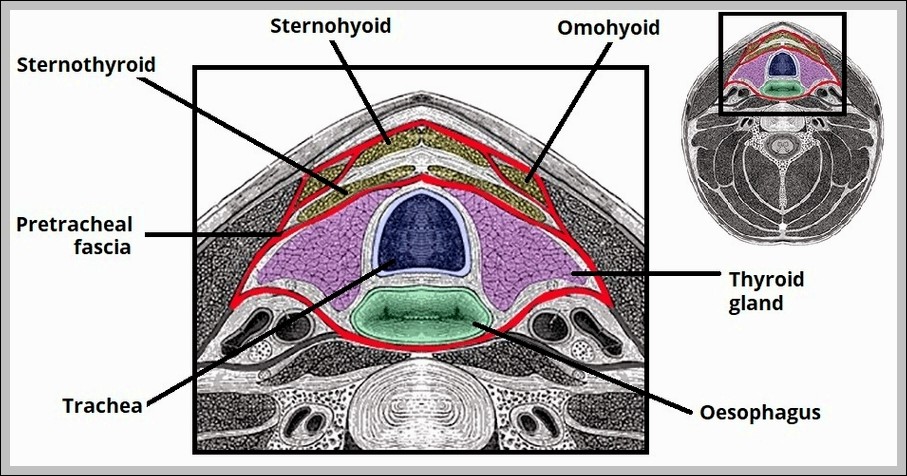
Pretracheal fascia (visceral layer) invests thyroid, trachea, esophagus, infrahyoid muscles; blends with carotid sheath laterally, buccopharyngeal fascia posteriorly. It forms visceral vascular space and invests recurrent laryngeal nerves.