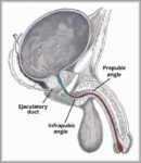
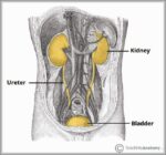
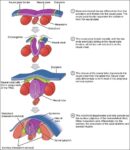
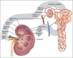
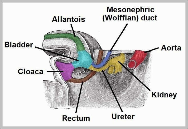
Upper Extremity Diagram

The forearm is the portion between the elbow and wrist. The thigh is the portion of the lower extremity between the hip and knee, and the calf is the portion between the knee and ankle. The normal arterial anatomy of the upper extremity is depicted graphically in Figure 13-1.
Muscles of the Upper Extremity. The pectoralis major, latissimus dorsi, deltoid, and rotator cuff muscles connect to the humerus and move the arm. The muscles that move the forearm are located along the humerus, which include the triceps brachii, biceps brachii, brachialis, and brachioradialis.
Use these carpal bones quizzes to remember carpal bones for good! Read article The lower extremity is a region of the body containing the hip, thigh, knee, leg, ankle and foot, all of which enable us to perform movements like walking, jumping and running. Start learning about these structures right away our free quiz guides below.

Upper Extremity Diagram Diagram - Upper Extremity Diagram Chart - Human anatomy diagrams and charts explained. This anatomy system diagram depicts Upper Extremity Diagram with parts and labels. Best diagram to help learn about health, human body and medicine.

upper extremity diagram

Trunk Muscles Diagram

Ventral trunk muscles (overview) The trunk (torso) is the central part of the body to which the head and the limbs are attached. Except for the brain, the trunk houses all the vital organs of the human body. The torso muscles attach to the skeletal core of the trunk, and depending on their location are divided into two large groups:
The trunk (torso) is the central part of the body to which the head and the limbs are attached. Except for the brain, the trunk houses all the vital organs of the human body. The torso muscles attach to the skeletal core of the trunk, and depending on their location are divided into two large groups:
The trunk (torso) is the central part of the body to which the head and the limbs are attached. Except for the brain, the trunk houses all the vital organs of the human body.

Trunk Muscles Diagram Diagram - Trunk Muscles Diagram Chart - Human anatomy diagrams and charts explained. This anatomy system diagram depicts Trunk Muscles Diagram with parts and labels. Best diagram to help learn about health, human body and medicine.

trunk muscles diagram

Tonsils Diagram

sagittal view of tonsils and throat anatomy. [edit on Wikidata] Tonsils are collections of lymphoid tissue facing into the aerodigestive tract. The set of lymphatic tissue known as Waldeyer’s tonsillar ring includes the adenoid tonsil, two tubal tonsils, two palatine tonsils, and the lingual tonsil.
The palatine tonsils and the adenoid tonsil are organs consisting of lymphoepithelial tissue located near the oropharynx and nasopharynx (parts of the throat). Humans are born with four types of tonsils: the pharyngeal tonsil, two tubal tonsils, two palatine tonsils and the lingual tonsils.
Tonsil Conditions. Acute tonsillitis: A bacteria or virus infects the tonsils, causing swelling and a sore throat. The tonsil may develop a gray or white coating (exudate). Chronic tonsillitis: Persistent infection of the tonsils, sometimes as a result of repeated episodes of acute tonsillitis.

Tonsils Diagram Diagram - Tonsils Diagram Chart - Human anatomy diagrams and charts explained. This anatomy system diagram depicts Tonsils Diagram with parts and labels. Best diagram to help learn about health, human body and medicine.

tonsils diagram

Thyroid Parathyroid Diagram

The parathyroid glands are a collection of four round glands around the size of a grain of rice. These glands are attached to the posterior surface of the lateral lobes of the thyroid gland.
It wraps around the anterior portion of the trachea and expands significantly on the lateral sides of the trachea and larynx. The lateral masses are known as the left and right lateral lobes, while the narrower medial region is known as the isthmus. The parathyroid glands are a collection of four round glands around the size of a grain of rice.
Normally, the thyroid is a deep brown color and the parathyroid adenoma is a mustard yellow color. Even though the colors are not well preserved, you can easily see the large round parathyroid adenoma completely within the thyroid gland.

Thyroid Parathyroid Diagram Diagram - Thyroid Parathyroid Diagram Chart - Human anatomy diagrams and charts explained. This anatomy system diagram depicts Thyroid Parathyroid Diagram with parts and labels. Best diagram to help learn about health, human body and medicine.

thyroid parathyroid diagram

Thymus Diagram

The thymus gland is a small organ behind the breastbone that plays an important function both in the immune system and endocrine system.
The growth and development of thymus continues until puberty. There are two distinct cell types within the thymus i.e. the lymphoid cells (thymocytes) and the reticular epithelial cells. In children, T cells densely populate the cortex of the thymus. As the T cells develop they pass to the medulla, before they are released into the circulation.
Within the thymus, T cells mature. T cells are critical to the adaptive immune system, where the body adapts specifically to foreign invaders. The thymus is composed of two identical lobes and is located anatomically in the anterior superior mediastinum, in front of the heart and behind the sternum.

Thymus Diagram Diagram - Thymus Diagram Chart - Human anatomy diagrams and charts explained. This anatomy system diagram depicts Thymus Diagram with parts and labels. Best diagram to help learn about health, human body and medicine.

thymus diagram

Thoracic Cage Diagram

The angles of the ribs form the most posterior extent of the thoracic cage. In the anatomical position, the angles align with the medial border of the scapula. A shallow costal groove for the passage of blood vessels and a nerve is found along the inferior margin of each rib. The bony ribs do not extend anteriorly completely around to the sternum.
The thoracic cage consists of ribs and the sternum. It is a bony structure that protects the organs in the thoracic cavity. It includes 25 bones, i.e., 1 sternum and 24 ribs. A. Ribs: They are a cage-like formation of thin, flat and curved bones.
The potential for movement is related to the flexibility provided by the ribs and their joints. The thoracic cage is composed of the thoracic skeleton, which includes the sternum, 12 pairs of ribs and 12 thoracic vertebrae, associated with the costal cartilages and intervertebral discs, respectively.

Thoracic Cage Diagram Diagram - Thoracic Cage Diagram Chart - Human anatomy diagrams and charts explained. This anatomy system diagram depicts Thoracic Cage Diagram with parts and labels. Best diagram to help learn about health, human body and medicine.

thoracic cage diagram

Testis Diagram

The testes or testicles are part of the male genitalia or sexual reproductive organ. It is a structure that is ovoid in shape and between four to six centimeters in length. These bilateral though not always symmetrical anatomical structures are the site of sperm production.
Learn about testicular anatomy, the functions of the testes, what takes place in between the seminiferous tubules, and the importance of the testes in human reproduction. Updated: 08/23/2021 The testes is where it all begins – sperm production, that is.
The testes are connected to the vital organs of the ventral body cavity via the spermatic cords. Nerves, blood vessels, and lymphatic vessels travel through the spermatic cords to support the testes. The vas deferens also passes through the spermatic cord carrying sperm out of the testes toward the prostate and urethra.

Testis Diagram Diagram - Testis Diagram Chart - Human anatomy diagrams and charts explained. This anatomy system diagram depicts Testis Diagram with parts and labels. Best diagram to help learn about health, human body and medicine.

testis diagram

Testis (2) Diagram

The testes are covered by outer fibrous tunica vaginalis, middle tunica albuginea, and inner tunica vasculosa. Internally, the testes are divided into about 250-300 pyramid-shaped compartments called testicular lobules.
On the testis, we can observe two sides (medial and lateral) that are separated by two edges (anterior and posterior). We can also observe superior and inferior poles since it is an ovoid organ.
The testes have two primary functions. The first function of the testes is to produce testosterone. Testosterone is a steroid hormone that is responsible for sexual differentiation of the male. It is important for prenatal development.

Testis (2) Diagram Diagram - Testis (2) Diagram Chart - Human anatomy diagrams and charts explained. This anatomy system diagram depicts Testis (2) Diagram with parts and labels. Best diagram to help learn about health, human body and medicine.

testis 2 diagram

Systemic Circuit Diagram

The systemic circuit is that part of your circulatory system that carries blood away from your heart, delivers it to most of your organs and tissues, and returns it to your heart again. The systemic circuit is distinct from the pulmonary circuit, which only conducts blood between your heart and lungs.
A schematic circuit diagram represents the electrical system in the form of a picture that shows the main features or relationships but not the details. In a schematic circuit diagram, the presentation of electrical components and wiring does not entirely correspond to the physical arrangements in the real device.
The blood vessels of the body are functionally divided into two distinctive circuits: pulmonary circuit and systemic circuit. The pump for the pulmonary circuit, which circulates blood through the lungs, is the right ventricle. The left ventricle is the pump for the systemic circuit, which provides the blood supply for the tissue cells of the body.

Systemic Circuit Diagram Diagram - Systemic Circuit Diagram Chart - Human anatomy diagrams and charts explained. This anatomy system diagram depicts Systemic Circuit Diagram with parts and labels. Best diagram to help learn about health, human body and medicine.

systemic circuit diagram

Synovial Joint Diagram

It is the most common type of joint found in the human body, and contains several structures which are not seen in fibrous or cartilaginous joints. In this article we shall look at the anatomy of a synovial joint – the joint capsule, neurovascular structures and clinical correlations.
The skeletal system has a number of different. A synovial joint, also known as diarthrosis, joins bones with a fibrous joint capsule that is continuous with the periosteum of the joined bones, constitutes the.Synovial Joint Labelled Diagram is free HD wallpaper. This wallpaper was upload at March 25, upload by admin in Diagram System.
At synovial joints, the articular surfaces of bones are covered with smooth articular cartilage. This gives the bones of a synovial joint the ability to move smoothly against each other, allowing for increased joint mobility. Figure 9.4.1 – Synovial Joints: Synovial joints allow for smooth movements between the adjacent bones.

Synovial Joint Diagram Diagram - Synovial Joint Diagram Chart - Human anatomy diagrams and charts explained. This anatomy system diagram depicts Synovial Joint Diagram with parts and labels. Best diagram to help learn about health, human body and medicine.

synovial joint diagram

Stomach Diagram

Given below is a labeled diagram of the stomach to help you understand stomach anatomy. The stomach is divided into four parts. These include: Cardia refers to the section of the stomach that is located around the cardiac orifice. The lower esophageal sphincter lies at the junction where the esophagus meets the stomach.
The diagram of the human digestive system is useful for both Class 10 and 12. It is one among the few important topics, which are repetitively asked in the board examinations. The diagram below shows the structure and functions of the human digestive system.
The stomach is a J-shaped organ that digests food. It produces enzymes (substances that create chemical reactions) and acids (digestive juices). This mix of enzymes and digestive juices breaks down food so it can pass to your small intestine. Your stomach is part of the gastrointestinal (GI) tract.

Stomach Diagram Diagram - Stomach Diagram Chart - Human anatomy diagrams and charts explained. This anatomy system diagram depicts Stomach Diagram with parts and labels. Best diagram to help learn about health, human body and medicine.

stomach diagram

Spleen Diagram

Spleen histology slide (labeled) The spleen is a fist sized organ located in the left upper quadrant of the abdomen. It is the largest lymphoid organ and thus the largest filter of blood in the human body. The spleen has a unique location, embryological development and histological structure that differs significantly from other lymphoid organs.
The spleen is an organ in the upper far left part of the abdomen, to the left of the stomach. The spleen varies in size and shape between people, but it’s commonly fist-shaped, purple, and about 4 inches long. … It acts as a filter for blood as part of the immune system.
The spleen is a purple, fist-sized organ. It is wrapped by a fibroelastic capsule which allows the spleen to significantly increase its size when necessary. The spleen is an intraperitoneal organ, so all of its surfaces are covered with visceral peritoneum.

Spleen Diagram Diagram - Spleen Diagram Chart - Human anatomy diagrams and charts explained. This anatomy system diagram depicts Spleen Diagram with parts and labels. Best diagram to help learn about health, human body and medicine.

spleen diagram

Reproductive Male Diagram

The male reproductive system consists of a pair of the testis, the duct system, accessory glands, and external genitalia. Let us understand in detail the male reproductive system through the diagram given below: The testicles are oval and located outside the abdominal cavity within a scrotum pouch.
Unlike Female Reproductive System, the male reproductive system is an external body part. Apart from these components, the male reproductive system also includes seminal vesicles, prostate glands, and bulbourethral glands, which perform various functions in the body.
Sexual reproduction is a biparental type of reproduction that involves the production of gametes and there is a fusion of male and female gametes to produce a diploid zygote. What is Male Reproductive System? The human male reproductive system is a group of organs that takes part in the process of reproduction and produces gametes.

Reproductive Male Diagram Diagram - Reproductive Male Diagram Chart - Human anatomy diagrams and charts explained. This anatomy system diagram depicts Reproductive Male Diagram with parts and labels. Best diagram to help learn about health, human body and medicine.

reproductive male diagram

Reproductive Female Diagram

The female reproductive system is composed of a pair of ovaries along with oviducts, vagina, cervix, uterus, and the external genitalia that are located in the pelvic region.
Female anatomy refers to the internal and external structures of the reproductive and urinary systems. Reproductive anatomy aids with sexual pleasure, getting pregnant, and breastfeeding a baby. The urinary system helps rid the body of toxins through urination (peeing).
What do the reproductive organs look like (picture)? The uterus, or womb, is a hollow organ located centrally in the pelvis. It houses the developing fetus during pregnancy. The lower portion of the uterus is called the cervix and opens into the vagina, or birth canal.

Reproductive Female Diagram Diagram - Reproductive Female Diagram Chart - Human anatomy diagrams and charts explained. This anatomy system diagram depicts Reproductive Female Diagram with parts and labels. Best diagram to help learn about health, human body and medicine.

reproductive female diagram

Pulmonary Circuit Diagram

Pulmonary circuit: Diagram of pulmonary circulation. Oxygen-rich blood is shown in red; oxygen-depleted blood in blue. Systemic circulation is the movement of blood from the heart through the body to provide oxygen and nutrients to the tissues of the body while bringing deoxygenated blood back to the heart.
Pulmonary Circuit. Pulmonary circulation transports oxygen-poor blood from the right ventricle to the lungs, where blood picks up a new blood supply. Then it returns the oxygen-rich blood to the left atrium.
Example Question #1 : Pulmonary And Systemic Circuits. The right atrium and ventricle transfer deoxygenated blood to the lungs via the pulmonary arteries. Blood is oxygenated and returned to the left artium via the pulmonary veins. The left ventricle then pumps the oxygenated blood to the body, exiting the heart through the aorta.

Pulmonary Circuit Diagram Diagram - Pulmonary Circuit Diagram Chart - Human anatomy diagrams and charts explained. This anatomy system diagram depicts Pulmonary Circuit Diagram with parts and labels. Best diagram to help learn about health, human body and medicine.

pulmonary circuit diagram

Planes Diagram

You might find a V-g diagram in your flight manual if you are flying a high performance fighter. The V-g diagram leads you to cornering speed and that allows you to extract maximum performance from your aircraft without breaking it.
A plane is an imaginary flat surface passing through the body or organ which divides the structure. 1) Frontal (Coronal) Plane: is vertical and extends from one side of the body to the other.
Airplanes are transportation devices which are designed to move people and cargo from one place to another. Airplanes come in many different shapes and sizes depending on the mission of the aircraft. The airplane shown on this slide is a turbine-powered airliner which has been chosen as a representative aircraft.

Planes Diagram Diagram - Planes Diagram Chart - Human anatomy diagrams and charts explained. This anatomy system diagram depicts Planes Diagram with parts and labels. Best diagram to help learn about health, human body and medicine.

planes diagram

Pituitary Pineal Glands Diagram

Pineal and Pituitary Glands. The pineal gland is a small endocrine gland in the brain, situated beneath the back part of the corpus callosum, and secretes melatonin. The pituitary gland is located at the base of the brain that secretes hormones and regulates and controls other hormone -secreting glands and many body processes,…
The pituitary gland is a pea-sized structure located at the base of the brain, just below the hypothalamus, to which it is attached via nerve fibers. It is part of the endocrine system and produces critical hormones, which are chemical substances that control various bodily functions.
Three glands of critical importance to the body, the hypothalamus, pituitary and pineal glands, are all located in the brain. These glands work synergistically to control many functions in the body.

Pituitary Pineal Glands Diagram Diagram - Pituitary Pineal Glands Diagram Chart - Human anatomy diagrams and charts explained. This anatomy system diagram depicts Pituitary Pineal Glands Diagram with parts and labels. Best diagram to help learn about health, human body and medicine.

pituitary pineal glands diagram

Pharynx Diagram

Anatomy of the pharynx (throat). The pharynx is a hollow tube that starts behind the nose, goes down the neck, and ends at the top of the trachea and esophagus. The three parts of the pharynx are the nasopharynx, oropharynx, and hypopharynx.
Laryngeal pharynx – It is the lowermost part of the pharynx from epiglottis and continues to the oesophagus. The main features of pharynx are: The wall of pharynx consists of both longitudinal and circular muscles. These muscles determine the shape of the lumen. Isthmus connects oropharynx to nasopharynx.
The Pharynx. The pharynx is a muscular tube that connects the oral and nasal cavity to the larynx and oesophagus. It begins at the base of the skull, and ends at the inferior border of the cricoid cartilage (C6).

Pharynx Diagram Diagram - Pharynx Diagram Chart - Human anatomy diagrams and charts explained. This anatomy system diagram depicts Pharynx Diagram with parts and labels. Best diagram to help learn about health, human body and medicine.

pharynx diagram

Penis Diagram

Penis Diagram Diagram - Penis Diagram Chart - Human anatomy diagrams and charts explained. This anatomy system diagram depicts Penis Diagram with parts and labels. Best diagram to help learn about health, human body and medicine.

penis diagram

Pelvic Girdle Diagram

The pelvic girdle, consisting of two hip bones, serves to attach the lower limbs to the sacrum of the axial skeleton. The right and left hip bones converge anteriorly and articulate with each other at the pubic symphysis. The combination of the two hip bones, the sacrum, and the coccyx forms the pelvis. The pelvis has a pronounced anterior tilt.
The wing of the ischium merges with the lateral surface of the ilium without any distinct boundary. The pubis of a chicken is the most ventral part of the chicken pelvic girdle. This is a thin elongated bone that incompletely fuses with the ischium bone of the chicken.
Pelvis, also called bony pelvis or pelvic girdle, in human anatomy, basin-shaped complex of bones that connects the trunk and the legs, supports and balances the trunk, and contains and supports the intestines, the urinary bladder, and the internal sex organs. The pelvis consists of paired hipbones, connected in front at the pubic symphysis and …

Pelvic Girdle Diagram Diagram - Pelvic Girdle Diagram Chart - Human anatomy diagrams and charts explained. This anatomy system diagram depicts Pelvic Girdle Diagram with parts and labels. Best diagram to help learn about health, human body and medicine.

pelvic girdle diagram