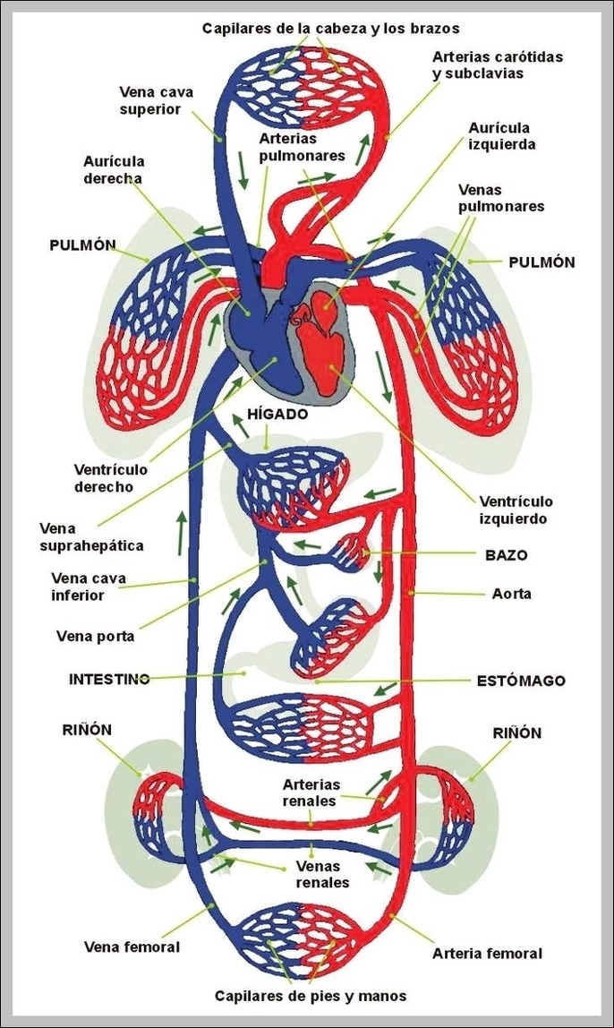
The inferior vena cava (also known as IVC or the posterior vena cava) is a large vein that carries blood from the torso and lower body to the right side of the heart.
Superior vena cava – ventral view. There is no valve that divides the SVC from the right atrium, which conducts blood from right atrial and right ventricular contractions upwards into the internal jugular vein (seen as the jugular venous pressure) and sternocleidomastoid muscle.
Superior vena cava – ventral view. There is no valve that divides the SVC from the right atrium, which conducts blood from right atrial and right ventricular contractions upwards into the internal jugular vein (seen as the jugular venous pressure) and sternocleidomastoid muscle.
Posterior Vena Cava Image
Posted inDiagrams
Posterior Vena Cava Image
Post navigation
Previous Post
 Picture Of The Internal Human Body Image
Next Post
Biomed Tech Schools Image