Types Of Muscle Tissue In Human Body Explained

Types of Muscle Tissue in the Human Body

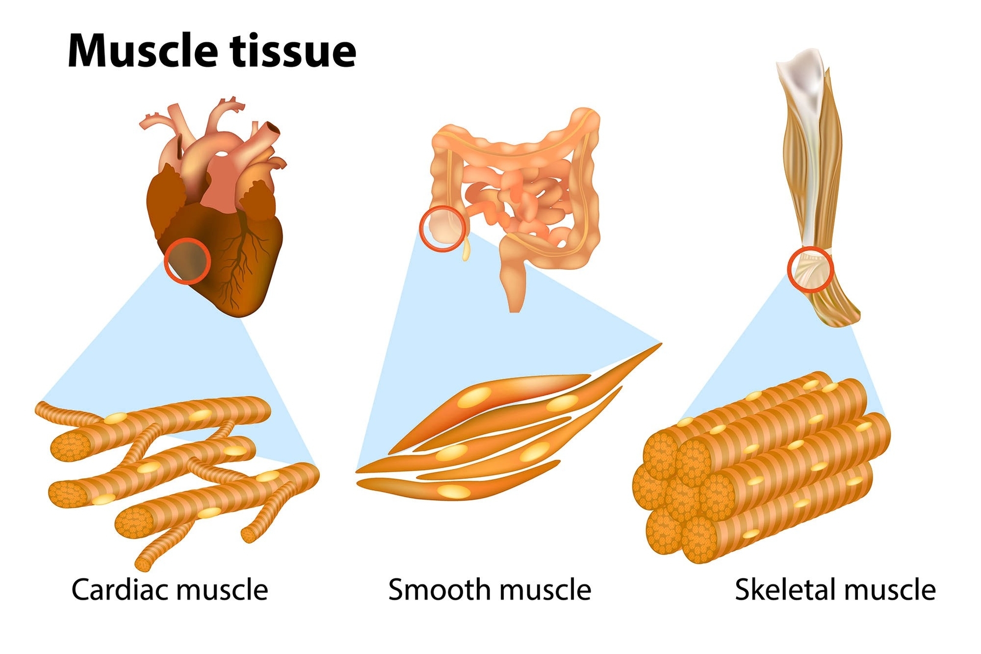
Muscle tissue is a specialized tissue found in animals which functions by contracting, thereby applying forces to different parts of the body. There are three types of muscle tissue: skeletal, cardiac, and smooth.

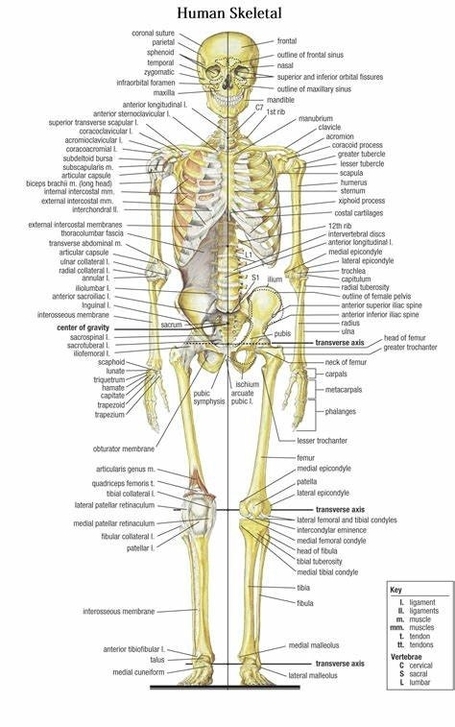
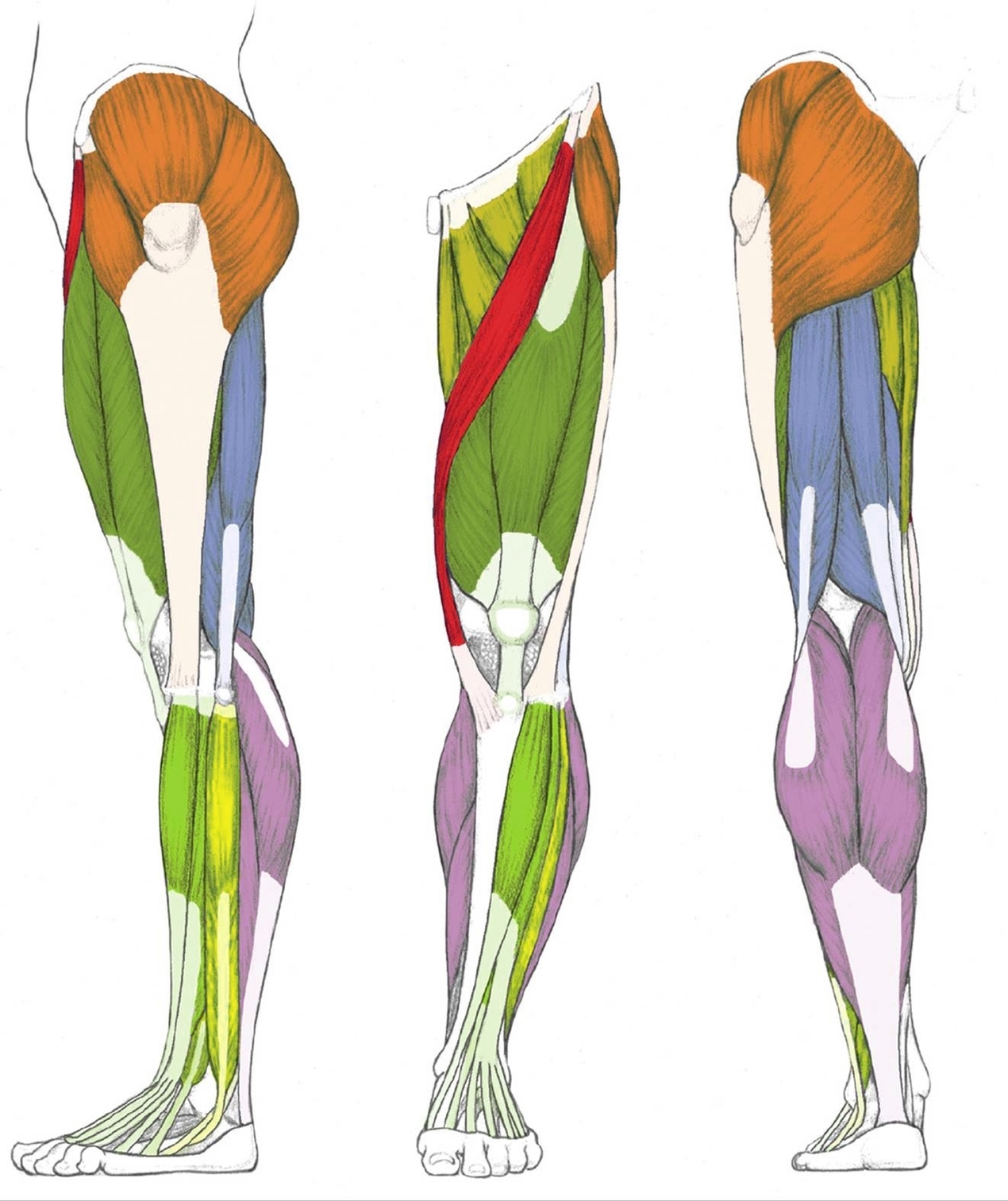
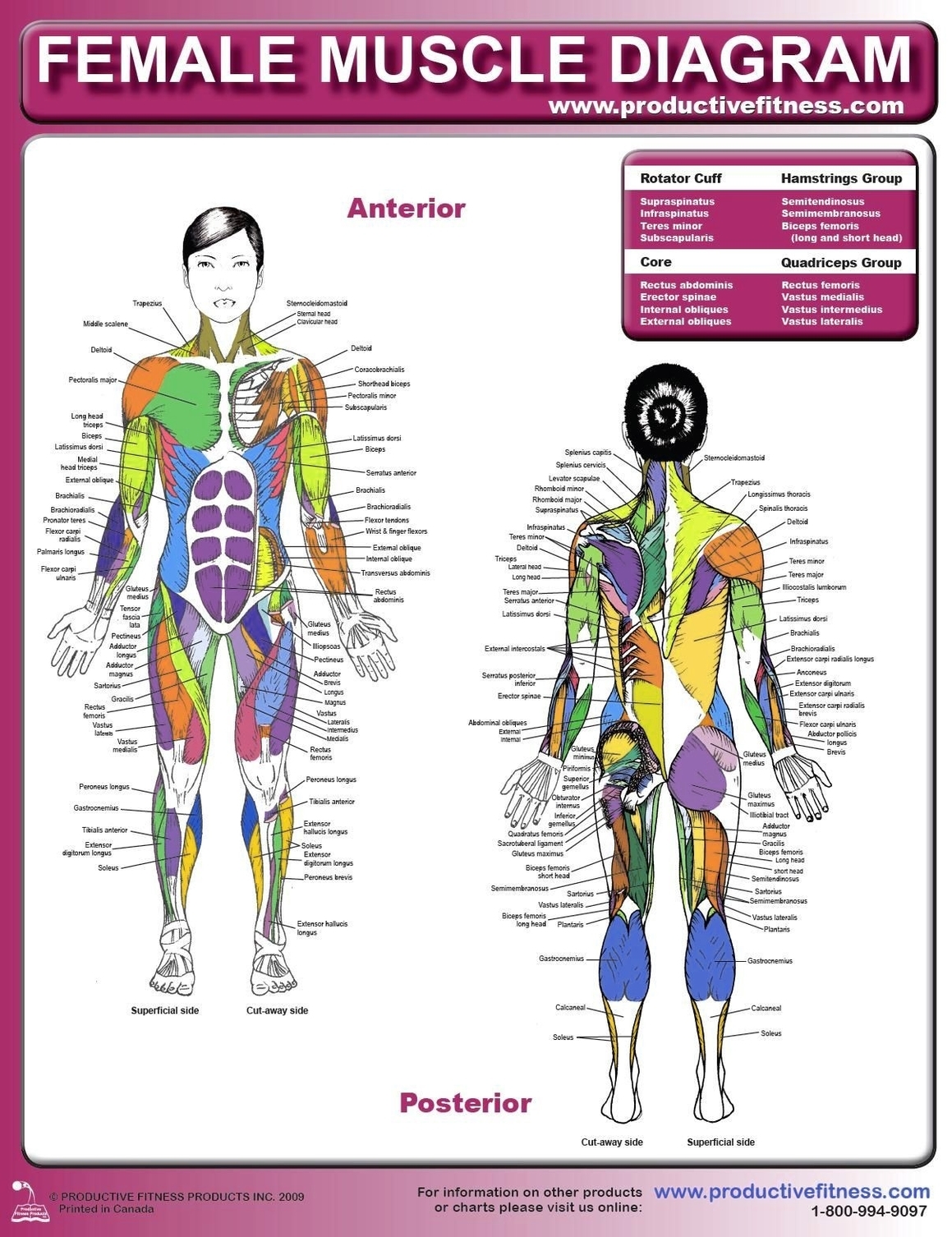
1. Skeletal Muscle: This type of tissue is found in skeletal muscles and is responsible for the voluntary movements of bones. It is a type of striated muscle, meaning clear bands can be seen in it under a microscope. These tiny light and dark bands are highly organized bundles of actin, myosin, and associated proteins. These organized bundles allow striated muscle to contract quickly and release quickly. Skeletal muscle tissue can be controlled voluntarily, by the brain.

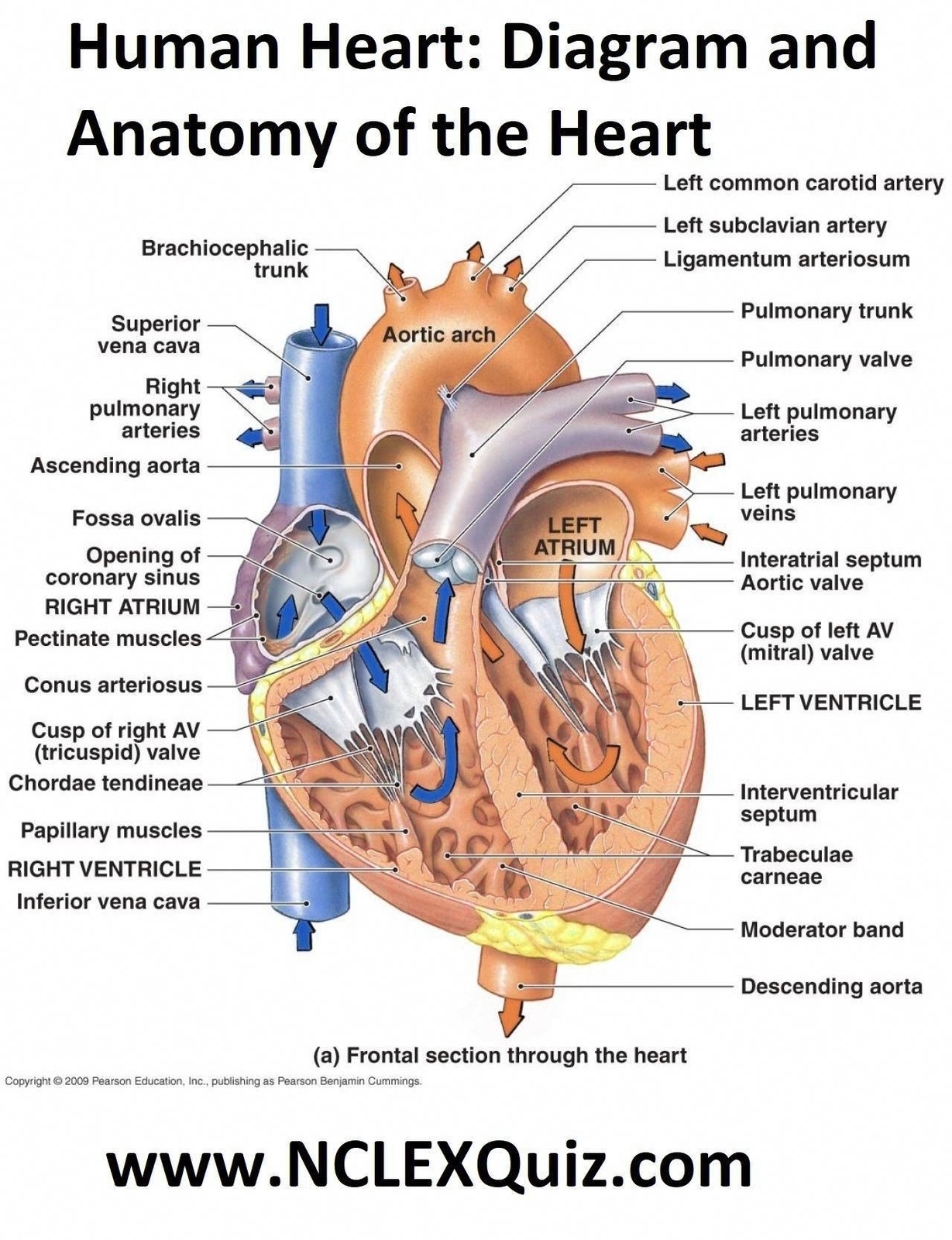
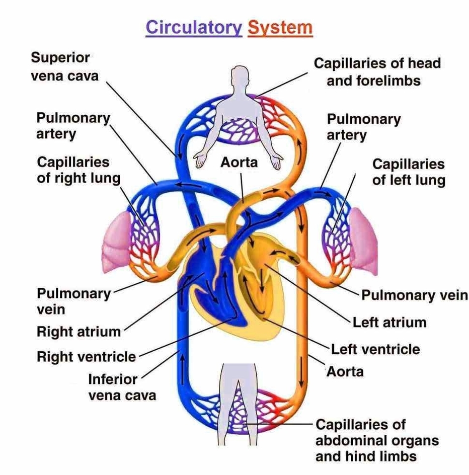
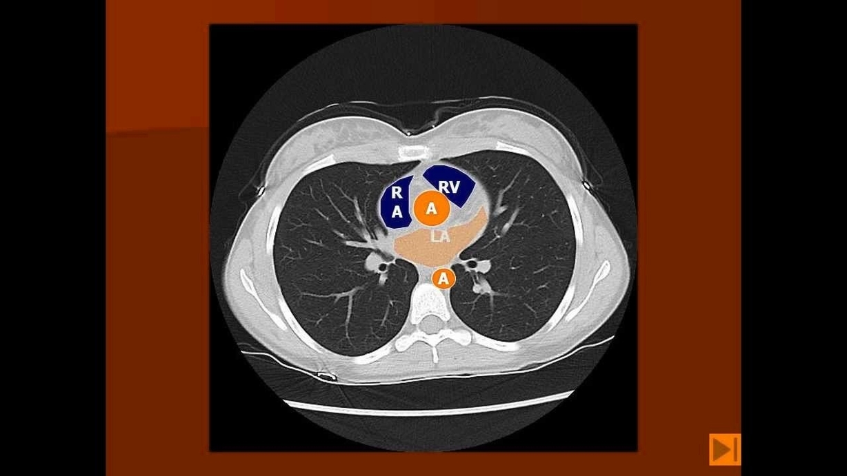
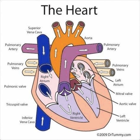
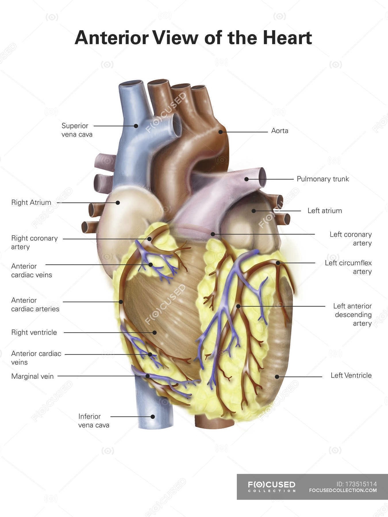
2. Cardiac Muscle: This is the specialized muscle of the heart. It is also a type of striated muscle, but unlike skeletal muscle, it is formed of short, uninucleate, branching myocytes which are connected at intercalated discs. These junctures help cardiac muscle to contract as one and provide a rapid and coordinated contraction to move blood.

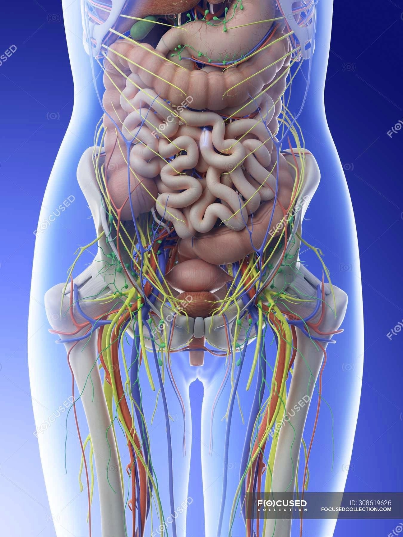
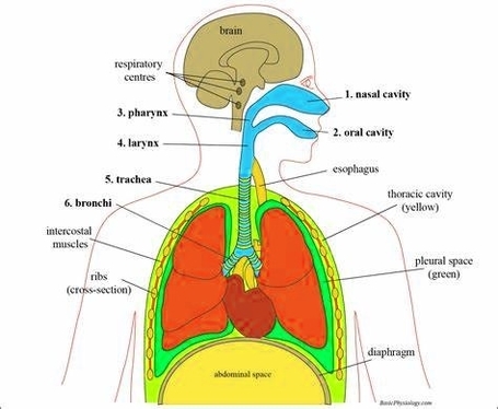
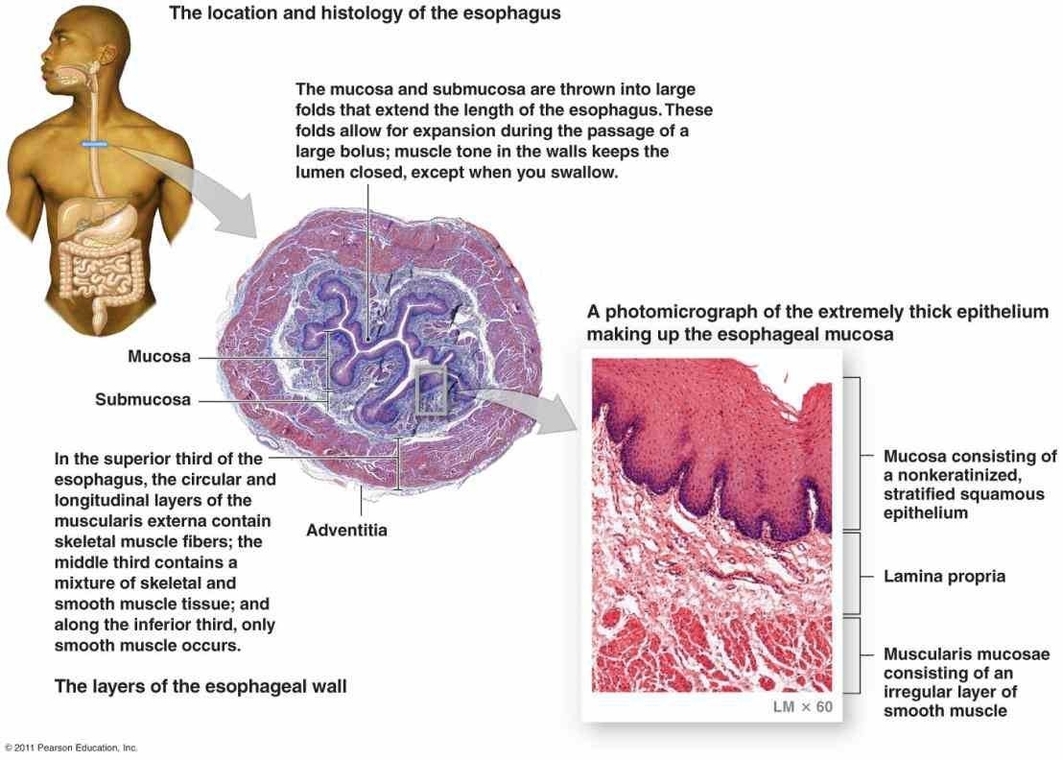
3. Smooth Muscle: This is a non-striated muscle, formed of short, uninucleate, spindle-shaped myocytes. It is located in the walls of internal organs, blood vessels, etc. Unlike cardiac and skeletal muscle tissue, smooth muscle tissue has no striations. The fibers of myosin and actin in smooth muscle fiber are not nearly as organized as in the other types of muscle tissue.

Each type of muscle tissue in the human body has a unique structure and a specific role. Skeletal muscle moves bones and other structures. Cardiac muscle contracts the heart to pump blood. Smooth muscle can be found in the walls of hollow organs, such as the intestines, uterus, and stomach.

Muscle tissue has four main properties: Excitability (an ability to respond to stimuli), Contractibility (an ability to contract), Extensibility (an ability to be stretched without tearing), and Elasticity (an ability to return to its normal shape). Through these properties, the muscular system as a whole performs several important functions. These include the production of force and movement, support of body stature and position, stability of joints, production of body heat (to maintain normal body temperature), as well as, provision of form to the body.

In conclusion, muscle tissues play a crucial role in the functioning of the human body. Their ability to contract and relax enables us to move, pump blood, and perform various other vital functions. Understanding the different types of muscle tissues and their functions can provide valuable insights into how our bodies work..

Types Of Muscle Tissue In Human Body Explained Diagram - Types Of Muscle Tissue In Human Body Explained Chart - Human anatomy diagrams and charts explained. This anatomy system diagram depicts Types Of Muscle Tissue In Human Body Explained with parts and labels. Best diagram to help learn about health, human body and medicine.

Types Of Muscle Tissue In Human Body Explained

Heart Diagram Coronary Sinus

The Coronary Sinus: An Overview

The coronary sinus (CS) is a significant component of the heart’s circulatory system. It is the largest vein of the heart, responsible for draining over half of the deoxygenated blood from the heart muscle into the right atrium. The CS plays a crucial role in life-saving heart treatments.

Anatomy and Location

The CS is a large coronary vein that measures between 3–5 centimeters in length and 1–2 cm in diameter. It is located towards the posterior, or rear, of the heart, between the left atrium and the left ventricle. The starting point of the CS is often described as where the great cardiac vein and the oblique vein of the left atrium meet. The large blood vessel then continues between the left atrium and left ventricle, running along the interventricular groove, and finally empties into the right atrium.

Function

The primary function of the CS is to drain deoxygenated blood from the heart muscle into the right atrium. This blood comes from most of the blood vessels of the heart muscle and wall. The CS is responsible for returning approximately 55% of the heart’s deoxygenated blood supply into the right atrium.

Tributaries

Many tributaries, or smaller veins, feed into and form the CS, though the exact anatomy may differ among individuals. Some veins that may feed into and form the CS include the great cardiac vein, oblique vein of the left atrium, posterior vein of the left ventricle, middle cardiac vein, and small cardiac vein.

Anatomical Variations

There are many different anatomical variations of the CS ranging from size, the number of smaller veins that connect to it, and the shape, or form, of the valves that surround it. Most anatomical variations are harmless, although some may present clinical implications for cardiac procedures.

Clinical Significance

The anatomical location of the CS often serves as a landmark for surgeons when performing cardiac surgery. The presence of irregular valves in the CS may hinder some cardiac intervention techniques.

Conclusion

The coronary sinus is a vital part of the heart’s circulatory system. Its primary function is to drain deoxygenated blood from the heart muscle into the right atrium. Understanding its anatomy, function, and clinical significance is crucial in the field of cardiology and cardiac surgery..

Heart Diagram Coronary Sinus Diagram - Heart Diagram Coronary Sinus Chart - Human anatomy diagrams and charts explained. This anatomy system diagram depicts Heart Diagram Coronary Sinus with parts and labels. Best diagram to help learn about health, human body and medicine.

Heart Diagram Coronary Sinus

Female Abdominal Anatomy And Internal Organs

The female abdominal anatomy is a complex and intricate system that houses various organs, each with its unique function. These organs are protected by the abdominal muscles, which include the rectus abdominis in front, the external obliques at the sides, and the latissimus dorsi muscles in the back.

Major Organs

1. Stomach, Small Intestine, and Large Intestine: These organs are responsible for digestion. They turn nutrients into usable energy and help dispose of solid waste.

2. Liver: Located in the upper right-hand part of the abdominal cavity, under the ribs, the liver processes blood, separating waste from nutrients.

3. Gallbladder: This tiny sack under the liver holds extra bile made by the liver until it is pumped into the small intestine. Bile helps break down fat.

4. Pancreas: This gland produces enzymes to help your body digest proteins, carbohydrates, and fats. It also makes hormones that help regulate the distribution of nutrients, including sugar.

5. Kidneys: Located near the back of the body, under the ribs, on each side of the spine, kidneys filter waste out of the bloodstream, which is passed out of the body as urine. They also help regulate levels of electrolytes, like salt and potassium, and produce certain hormones.

6. Suprarenal (Adrenal) Glands: These glands synthesize and secrete hormones that help the kidneys conserve sodium, thus conserving water. They also play a role in supporting the body’s sexual functions.

Female Reproductive Organs

1. Uterus (Womb): A hollow, pear-shaped organ located in a woman’s lower abdomen, between the bladder and the rectum.

2. Ovaries: Two female reproductive organs located in the pelvis.

3. Fallopian Tubes: These carry eggs from the ovaries to the uterus.

4. Cervix: The lower part of the uterus that opens into the vagina.

5. Vagina: The canal that joins the cervix to the outside of the body.

External Female Anatomy

1. Mons Pubis: The rounded, fleshy area on the front of the pelvic bone where pubic hair usually grows.

2. Labia Majora and Minora: The fleshy outer and inner folds of protective skin located on each side of the vaginal opening.

3. Clitoris: Sits at the top of the vulva, above the urethral opening.

4. Urethral Opening: The tube that carries urine from the bladder to the outside of the body.

These organs and structures work together to perform a variety of functions, including digestion, waste removal, reproduction, and hormone regulation. Understanding the female abdominal anatomy is crucial for maintaining health and diagnosing potential health issues..

Female Abdominal Anatomy And Internal Organs Diagram - Female Abdominal Anatomy And Internal Organs Chart - Human anatomy diagrams and charts explained. This anatomy system diagram depicts Female Abdominal Anatomy And Internal Organs with parts and labels. Best diagram to help learn about health, human body and medicine.

Female Abdominal Anatomy And Internal Organs

Human Heart Diagram Illustrated Representation

The Human Heart: An Illustrated Representation

The human heart, a muscular organ roughly the size of a closed fist, serves as the body’s circulatory pump. It is located in the thoracic cavity, medial to the lungs and posterior to the sternum. The base of the heart is attached to the aorta, pulmonary arteries and veins, and the vena cava, while the apex points towards the left side.

Anatomy of the Heart

1. Pericardium: The heart resides within a fluid-filled cavity called the pericardial cavity. The pericardium, a serous membrane, lines this cavity, producing serous fluid to lubricate the heart and prevent friction.

2. Heart Wall: Comprising three layers—epicardium, myocardium, and endocardium—the heart wall encases the organ. The epicardium, also known as the visceral layer of the pericardium, forms the outermost layer.

3. Chambers and Valves: The heart consists of four chambers—two atria (left and right) and two ventricles (left and right). The atria receive blood, while the ventricles pump blood out. Four valves (aortic, mitral, tricuspid, and pulmonary) ensure unidirectional blood flow.

4. Blood Vessels: The heart connects to several blood vessels, including the aorta, pulmonary arteries and veins, and the vena cava.

5. Conduction System: This includes the sinuatrial node, atrioventricular node, and atrioventricular bundle, which regulate the heart’s rhythm.

Illustrated Anatomy

Illustrated representations of the heart, based on medical illustrations and cadaver photography, offer an interactive way to explore heart anatomy. These illustrations, often labeled, serve as invaluable medical and anatomical tools. They depict various anatomical structures, including the myocardium, valves, coronary arteries, and the conduction system.

3D Models

3D models provide a comprehensive view of the heart’s anatomy. They allow for rotation and zooming, offering a detailed perspective of different parts, such as the aortic valve, bundle branches, chordae tendineae, and the interventricular septum.

Conclusion

Understanding the human heart’s anatomy is crucial in the medical field. Illustrated representations and 3D models serve as effective educational tools, offering an interactive and detailed exploration of the heart’s complex structure.

Human Heart Diagram Illustrated Representation Diagram - Human Heart Diagram Illustrated Representation Chart - Human anatomy diagrams and charts explained. This anatomy system diagram depicts Human Heart Diagram Illustrated Representation with parts and labels. Best diagram to help learn about health, human body and medicine.

Human Heart Diagram Illustrated Representation

Decedent Estate Flowchart

Decedent Estate Flow

The process of managing and distributing a deceased person’s estate, often referred to as the “Decedent Estate Flow,” involves several key steps and legal considerations. Here’s an overview:

1. Decedent and Legal Rights: A decedent can control where their assets go after they die via a will or trust. They have legal and financial obligations, including distributing their assets and paying outstanding taxes or debts, which they carry out through representatives such as an executor, administrator, or trustee.

2. Wills and Estate Laws in Canada: Inheritance laws, also called wills and estate laws in Canada, govern the distribution of a person’s wealth when they die. These laws provide for minimum standards that wills should follow to be valid, court processes to be undertaken when the will’s validity is being questioned, minimum requisites in appointing a valid executor of the deceased’s will, an administrator if there’s no will, or a valid guardian for the deceased’s minor children.

3. Probate Process: Probate is a process that verifies the deceased’s last will and testament. If there wasn’t a will, then the probate process confirms who the executor of the estate can be. The probate process helps to clarify these things as certain steps, forms, and notifications must be completed.

4. Estate Expenses During Probate: An executor is expected to pay the expenses of an estate on time, but what happens if the estate is tied up in the probate process? This includes taxes on income from registered retirement savings plans (RRSPs).

5. Period for Settling an Estate: For taxation and other legal purposes, the estate of a deceased person must be settled after the issuance of the Grant of Letters Probate and within the period stated in provincial wills and estate laws.

6. Deceased with a Will: In common law provinces, when a person dies with a will, what is stated in the will must be followed by the family. This may cover aspects like the distribution of wealth and the succession of properties, among others.

In conclusion, the Decedent Estate Flow is a complex process that involves various legal and financial considerations. It’s crucial to understand these aspects to ensure the smooth transition of the decedent’s assets and to fulfill their final wishes.

Decedent Estate Flowchart Diagram - Decedent Estate Flowchart Chart - Human anatomy diagrams and charts explained. This anatomy system diagram depicts Decedent Estate Flowchart with parts and labels. Best diagram to help learn about health, human body and medicine.

Decedent Estate Flowchart

Human Bones Anatomy

Human Bones Anatomy

The human skeleton, an internal framework, is composed of individual bones and cartilages. It’s intimately associated with fibrous connective tissues, the ligaments, and tendons. The skeleton can be divided into two main parts:

1. Axial Skeleton: Comprises the vertebral column, much of the skull, and the visceral part, which includes the lower jaw, some elements of the upper jaw, and the branchial arches, including the hyoid bone.
2. Appendicular Skeleton: Includes the pelvic (hip) and pectoral (shoulder) girdles and the bones and cartilages of the limbs.

The skeleton performs three primary functions: support, protection, and motion. The vertebral column, corresponding to the notochord in lower organisms, is the main support of the trunk. The central nervous system lies largely within the axial skeleton, the brain being well protected by the cranium and the spinal cord by the vertebral column.

Bones are living tissues that make up the body’s skeleton. There are three types of bone tissue:

1. Compact Tissue: The harder, outer tissue of bones.
2. Cancellous Tissue: The sponge-like tissue inside bones.
3. Subchondral Tissue: The smooth tissue at the ends of bones, covered with cartilage.

The tough, thin outer membrane covering the bones is called the periosteum. Beneath the periosteum’s hard outer shell are tunnels and canals through which blood and lymphatic vessels run to carry nourishment for the bone. Muscles, ligaments, and tendons may attach to the periosteum.

Bones are classified by their shape—as long, short, flat, and irregular. Primarily, they are referred to as long or short. The adult human skeleton consists of 206

Human Bones Anatomy Diagram - Human Bones Anatomy Chart - Human anatomy diagrams and charts explained. This anatomy system diagram depicts Human Bones Anatomy with parts and labels. Best diagram to help learn about health, human body and medicine.

Human Bones Anatomy

Normal Chest Ct With Labels

A Chest Computed Tomography (CT) is a non-invasive diagnostic imaging procedure that uses special X-ray equipment to create detailed pictures, or scans, of areas inside the body. It is also sometimes referred to as a CAT scan (Computed Axial Tomography). It is used to visualize the heart, lungs, blood vessels, and bones in the chest region.

A normal chest CT scan reveals several key features:

1. Lungs and Airways: The lungs and airways appear normal in size and shape without any signs of inflammation. In a healthy lung, the diaphragm appears domed.

2. Pleura: The pleura, the thin membrane that lines the outer surface of the lungs and the inner surface of the chest wall, shows no signs of effusion (fluid) or thickening.

3. Heart: The heart size is normal without any signs of enlargement.

4. Pericardium: The pericardium, the double-walled sac containing the heart and the roots of the great vessels, shows no signs of effusion (fluid).

5. Mediastinum: The mediastinum, the central compartment of the thoracic cavity, and its structures have a normal configuration.

6. Chest Wall: The chest wall, which includes the ribs, muscles, and skin, is unremarkable.

A chest CT scan can be performed with or without contrast. Contrast is a special dye injected into the patient to make the structures inside the body more visible in the CT images. A normal CT chest with intravenous contrast is performed for a patient with sudden tearing central chest pain.

The rise of modern technology has allowed for the development of more advanced imaging techniques. For example, volumetric axial images can be obtained through the thorax without contrast media injection. These techniques provide a more detailed view of the chest’s anatomy, aiding in the diagnosis and treatment of various conditions.

In conclusion, a normal chest CT scan provides a detailed view of the chest’s anatomy, including the heart, lungs, blood vessels, and bones. It is a valuable tool in diagnosing and monitoring a wide range of health conditions. However, it is important to note that while a CT scan provides detailed images of the chest, it is only one piece of the puzzle. A comprehensive understanding of a patient’s health requires a combination of imaging results, physical examination, and medical history..

Normal Chest Ct With Labels Diagram - Normal Chest Ct With Labels Chart - Human anatomy diagrams and charts explained. This anatomy system diagram depicts Normal Chest Ct With Labels with parts and labels. Best diagram to help learn about health, human body and medicine.

Normal Chest Ct With Labels

Human Leg Muscles Described

The human leg, a complex structure that enables movement and supports the body’s weight, is composed of numerous muscles. These muscles can be broadly categorized into those of the upper leg and those of the lower leg.

Upper Leg Muscles

The upper leg, or thigh, houses some of the body’s strongest muscles, including the quadriceps and hamstrings.

1. Quadriceps: These are the major extensors of the knee and are the strongest and leanest muscles in the body. They consist of four muscles:
– Vastus lateralis: The largest of the quadriceps, it extends from the top of the femur to the kneecap.
– Vastus medialis: A teardrop-shaped muscle of the inner thigh that attaches along the femur and down to the inner border of the kneecap.
– Vastus intermedius: Located between the vastus medialis and the vastus lateralis at the front of the femur, it is the deepest of the quadriceps muscles.
– Rectus femoris: This muscle attaches to the kneecap and has the least effect on flexion of the knee among the quadriceps muscles.

2. Hamstrings: These are three muscles at the back of the thigh that affect hip and knee movement. They include:
– Biceps femoris: A long muscle that flexes the knee.
– Semimembranosus: This long muscle extends from the pelvis to the tibia, extends the thigh, flexes the knee, and helps rotate the tibia.
– Semitendinosus: This muscle also extends the thigh and flexes the knee.

Lower Leg Muscles

The lower leg muscles, particularly the calf muscles, are pivotal to the movement of the ankle, foot, and toes.

1. Gastrocnemius (calf muscle): One of the large muscles of the leg, it connects to the heel and flexes and extends the foot, ankle, and knee.
2. Soleus: This muscle extends from the back of the knee to the heel and is important in walking and standing.
3. Plantaris: A small, thin muscle that is absent in about 10% of people.

The Achilles tendon, which connects the plantaris, gastrocnemius, and soleus muscles to the heel bone, is possibly the most important tendon in terms of mobility. It stores the elastic energy needed for running, jumping, and other physical activities.

In conclusion, the muscles of the human leg, working in harmony with bones, tendons, and ligaments, enable a wide range of movements, from walking and running to jumping and flexing the feet. They also play a crucial role in maintaining posture and supporting the body’s weight.

Human Leg Muscles Described Diagram - Human Leg Muscles Described Chart - Human anatomy diagrams and charts explained. This anatomy system diagram depicts Human Leg Muscles Described with parts and labels. Best diagram to help learn about health, human body and medicine.

Human Leg Muscles Described

Evolution Of The Human Heart Example Of

Evolution of the Human Heart

The human heart, a vital organ responsible for pumping blood throughout the body, is a product of millions of years of evolution. Its journey from a simple circulatory mechanism to a complex four-chambered structure is a fascinating tale of adaptation and survival.

Invertebrate Hearts

The earliest precursors to the human heart were found in invertebrate animals. Many of these creatures did not have a heart or blood, as they were not complex enough to require a system for nutrient delivery to their cells. As invertebrates became more complex, they developed an open circulatory system, where blood was pumped throughout the tissues and filtered back to the pumping mechanism.

Fish Hearts

The next step in the evolutionary chain is seen in fish, which have a two-chambered heart consisting of an atrium and a ventricle. This closed circulatory system has a single large vessel that carries blood to the gills for oxygenation and then transports it around the fish’s body.

Amphibian Hearts

Amphibians, like frogs, represent the link between aquatic and terrestrial animals. Frogs have a three-chambered heart with two atria and one ventricle. The separation of the atria allows frogs to keep oxygenated and deoxygenated blood separate as they enter the heart.

Reptile Hearts

Reptiles, such as turtles, have a heart that is a sort of three-and-a-half chambered heart. There is a small septum that goes about halfway down the ventricle. This change allows the turtle’s body to receive blood that is slightly richer in oxygen than that of a frog.

Human Hearts

The human heart, with its four chambers, is a result of further evolution. This configuration ensures the separation of low-pressure circulation to the lungs and high-pressure pumping into the rest of the body. The human heart has evolved over hundreds of thousands of years as our activity levels gradually became more sustained. It has spent millions of years perfecting itself to keep humans alive.

Conclusion

The evolution of the human heart is a testament to the power of natural selection and adaptation. From simple circulatory systems in invertebrates to the complex four-chambered heart in humans, each step in this journey has been driven by the need for survival and adaptation to changing environments. Today, the human heart is a marvel of biological engineering, capable of supporting our bodies’ needs in a wide range of conditions. Its evolution is a fascinating example of how life adapts and evolves over time..

Evolution Of The Human Heart Example Of Diagram - Evolution Of The Human Heart Example Of Chart - Human anatomy diagrams and charts explained. This anatomy system diagram depicts Evolution Of The Human Heart Example Of with parts and labels. Best diagram to help learn about health, human body and medicine.

Evolution Of The Human Heart Example Of

Scottish Organ Donors Illustration Description Example With Labels

Scottish Organ Donors

Organ donation in Scotland is a significant aspect of the country’s healthcare system. It involves the process of giving organs or tissues to help save or improve the lives of others who are critically ill. The system is managed by the NHS Organ Donor Register.

Opt-Out System
cotland operates an opt-out system for organ donation. This means that all adults in Scotland are automatically considered as potential donors unless they choose to opt out. The law changed to this system on March 26, 2021. However, medical staff will always consult the family about the individual’s latest views to ensure donation does not proceed if the person was against it.

Registration

Individuals have the option to register their decision about organ donation. They can choose to donate all or some types of organs and tissues after their death. Alternatively, they can register not to be a donor, which means opting out of the donation of all organs and tissues. It is recommended that individuals discuss their decision with family and friends.

Living Donation

In addition to posthumous organ donation, Scotland also recognizes living donation. This is particularly relevant for kidney donations, as there are currently over 400 people in Scotland in need of a kidney. Living kidney donation is an exceptional gift that can transform the life of someone on the transplant waiting list.

Impact of Donation

Organ donation has a profound impact on the lives of recipients. It not only provides a second chance at life for those who are critically ill but also improves the quality of life for many more. The stories of donor families and recipients highlight the life-changing impact of organ donation.

Conclusion

Organ donation in Scotland is a testament to the power of altruism and medical advancement. It is a personal decision that has far-reaching effects, offering hope and life to those in need. The opt-out system, while respecting individual choice, aims to increase the number of donors and thereby save more lives. As such, organ donation represents a vital part of Scotland’s healthcare landscape.

Scottish Organ Donors Illustration Description Example With Labels Diagram - Scottish Organ Donors Illustration Description Example With Labels Chart - Human anatomy diagrams and charts explained. This anatomy system diagram depicts Scottish Organ Donors Illustration Description Example With Labels with parts and labels. Best diagram to help learn about health, human body and medicine.

Scottish Organ Donors Illustration Description Example With Labels

Respiratory Systemrespiratory System In Animals

Respiratory System in Animals

The respiratory system in animals is a complex and vital system that facilitates the exchange of gases, primarily oxygen and carbon dioxide. It begins at the nose and ends at the distal alveoli.

Upper Airways

The upper airway includes the nose, sinuses, and pharynx. The nose provides olfaction and temperature regulation in hyperthermic patients. The nasal turbinates humidify and warm air, and filter particulate matter.

Lower Airways

The lower airways include the trachea, bronchi, bronchioles, and alveoli. The trachea is a stiff tube reinforced by rings of cartilage. It splits into two tubes called bronchi, which further split off many times to form smaller tubes called bronchioles. These bronchioles lead to sacs called alveoli.

Lungs

The lungs are a pair of respiratory organs used by mammals. They are comprised of millions of alveoli, where gas exchange occurs. Oxygen moves into the capillary from the alveoli, and carbon dioxide moves into the alveoli from the blood in the capillary.

Ventilation

Ventilation is the process by which air is pulled in and pushed out of the lungs. The diaphragm and intercostal muscles control the volume of the thoracic cavity, which houses the lungs. When you breathe in (inhalation), these muscles contract, increasing the volume of the thoracic cavity and causing air to rush into the lungs. When you breathe out (exhalation), these muscles relax, decreasing the space in the thoracic cavity and forcing air out of the lungs.

Defense Mechanisms

The respiratory system has several defense mechanisms. Large, inhaled airborne particles enter the nose and are deposited along the mucous lining of the nasal passages. Cilia move these particles along the mucosal barrier to the pharynx to be swallowed or expectorated. Small particles may be deposited in the alveoli, where they are phagocytized by macrophages.

Conclusion

The respiratory system is crucial for the survival of animals. It not only facilitates the exchange of gases but also plays a role in maintaining acid-base balance, acting as a blood reservoir, filtering and probably destroying emboli, metabolizing some bioactive substances, and activating some substances. Any major dysfunction of gas transfer due to disease can lead to respiratory distress or failure..

Respiratory Systemrespiratory System In Animals Diagram - Respiratory Systemrespiratory System In Animals Chart - Human anatomy diagrams and charts explained. This anatomy system diagram depicts Respiratory Systemrespiratory System In Animals with parts and labels. Best diagram to help learn about health, human body and medicine.

Respiratory Systemrespiratory System In Animals

Female Muscle Diagram And Definitions

Understanding Female Muscles and Definitions

Muscles are soft tissues found throughout the body, playing a crucial role in everything from maintaining posture to enabling movement. In females, muscle structure and function are influenced by various biological and hormonal factors.

Muscle Types and Functions

There are three types of muscle tissues in the body: skeletal, cardiac, and smooth.

1. Skeletal Muscles: These voluntary muscles are responsible for body movements and maintaining posture. They are attached to bones and contract and relax to facilitate movement.

2. Cardiac Muscles: Found only in the heart, these involuntary muscles contract rhythmically to pump blood throughout the body.

3. Smooth Muscles: These involuntary muscles are found in the walls of organs and structures such as the esophagus, stomach, intestines, bronchi, uterus, urethra, and blood vessels.

Female Muscle Structure and Growth

Women typically have a smaller skeletal structure than men, with a wider pelvis and thigh bones that come down at more of an angle. Women have about 90% of the potential leg mass and 50% of the upper body mass compared to men.

Women generally have lower levels of testosterone, a hormone critical for muscle building. This means women may not build muscle as quickly or to the same extent as men. Additionally, women tend to carry more body fat than men, which can make it more challenging to see visible muscle definition.

Role of Muscles in the Female Body

Muscles play a vital role in various bodily functions, including vision, hearing, breathing, speaking, swallowing, digesting food, eliminating waste, moving, sitting still, standing up straight, pumping blood, and giving birth. Muscles also store and release energy as part of metabolism.

Conclusion

Understanding the structure and function of muscles in the female body provides insight into the unique physiological characteristics of women. It’s important to note that while there are general differences between male and female muscle structure and growth, individual variations can be significant. Therefore, fitness and health strategies should be personalized to meet individual needs and goals.

Female Muscle Diagram And Definitions Diagram - Female Muscle Diagram And Definitions Chart - Human anatomy diagrams and charts explained. This anatomy system diagram depicts Female Muscle Diagram And Definitions with parts and labels. Best diagram to help learn about health, human body and medicine.

Female Muscle Diagram And Definitions

Cells Of The Digestive System

The digestive system, a complex network of organs, plays a crucial role in the body’s overall health and functioning. It is responsible for breaking down food into nutrients, which the body uses for energy, growth, and cell repair. The cells of the digestive system are specialized to perform specific functions that contribute to the process of digestion.
tomach Cells

The stomach, a muscular organ in the upper abdomen, contains various types of cells that secrete digestive enzymes and hydrochloric acid. These substances break down food so the body can absorb nutrients. The stomach’s cells are constantly exposed to digestive juices and the stomach’s contents. To protect themselves, the cells produce a layer of mucus and constantly regenerate to keep the stomach lining healthy.

1. Surface Mucous Cells: These cells secrete a thick layer of mucus that covers the stomach lining and protects it from the digestive juices. The mucus also provides a slippery surface that helps food move through the stomach.

2. Parietal Cells: These cells are present in the gastric pits that mainly occur in the upper part of the stomach, or the fundus. They secrete hydrochloric acid, which helps break down food. Hydrochloric acid also kills bacteria and other microorganisms that may be present in food.

Cells of the Gastrointestinal Tract

The gastrointestinal (GI) tract, which includes the mouth, esophagus, stomach, small intestine, large intestine, and anus, contains many specialized cells. These cells work together to ensure the smooth functioning of the digestive system.

1. Taste Cells: These cells are located in the mouth and are responsible for detecting the taste of food.

2. Pancreatic Duct Cells: These cells are part of the pancreas and play a role in the secretion of digestive enzymes.

3. Enterocytes: These cells line the small intestine and are responsible for the absorption of nutrients from digested food.

4. Microfold Cells: These cells are found in the lining of the intestines and play a role in the immune response of the gut.

Hormone-Secreting Cells

Cells lining your stomach and small intestine make and release hormones that control how your digestive system works. These hormones tell your body when to make digestive juices and send signals to your brain that you are hungry or full.

In conclusion, the cells of the digestive system are specialized to perform specific functions that contribute to the process of digestion. They work together to ensure the smooth functioning of the digestive system, playing crucial roles in breaking down food, absorbing nutrients, and maintaining overall health..

Cells Of The Digestive System Diagram - Cells Of The Digestive System Chart - Human anatomy diagrams and charts explained. This anatomy system diagram depicts Cells Of The Digestive System with parts and labels. Best diagram to help learn about health, human body and medicine.

Cells Of The Digestive System

Human Heart With Labels

The Human Heart

The human heart, a muscular organ roughly the size of a closed fist, is the epicenter of the circulatory system. It is located between the lungs, slightly to the left of center, behind the breastbone, and rests on the diaphragm.
tructure

The heart consists of several layers of a tough muscular wall, the myocardium. A thin layer of tissue, the pericardium, covers the outside, and another layer, the endocardium, lines the inside. The heart cavity is divided down the middle into a right and a left heart, which in turn are subdivided into two chambers. The upper chamber is called an atrium (or auricle), and the lower chamber is called a ventricle.

Function

The heart, although a single organ, can be considered as two pumps that propel blood through two different circuits. The right atrium receives venous blood from the head, chest, and arms via the large vein called the superior vena cava and receives blood from the abdomen, pelvic region, and legs via the inferior vena cava. Blood then passes through the tricuspid valve to the right ventricle, which propels it through the pulmonary artery to the lungs.

In humans, deoxygenated blood enters the heart through the right atrium from the superior and inferior venae cavae and passes to the right ventricle. From here, it is pumped into pulmonary circulation to the lungs, where it receives oxygen and gives off carbon dioxide. Oxygenated blood then returns to the left atrium, passes through the left ventricle and is pumped out through the aorta into systemic circulation.

Circulation

The heart powers the circulatory system, supplying the body with oxygen and other important nutrients needed to sustain life. The pumped blood carries oxygen and nutrients to the body, while carrying metabolic waste such as carbon dioxide to the lungs.

Heartbeat

The heart beats at a resting rate close to 72 beats per minute. The heart beats around 100,000 times a day, pumping approximately 8 pints of blood throughout the body 24/7. Exercise temporarily increases the rate, but lowers it in the long term, and is good for heart health.

Conclusion

The human heart, a marvel of biological engineering, is a testament to the intricate and efficient design of the human body. Its ceaseless work powers every function and process within our bodies, making it a symbol of life itself..

Human Heart With Labels Diagram - Human Heart With Labels Chart - Human anatomy diagrams and charts explained. This anatomy system diagram depicts Human Heart With Labels with parts and labels. Best diagram to help learn about health, human body and medicine.

Human Heart With Labels

Parts Of The Brain Diagram And Function

The human brain, the most complex organ in the body, is responsible for controlling thought, memory, emotion, touch, motor skills, vision, breathing, temperature, hunger, and every process that regulates our body. It is made up of billions of neurons and has several specialized parts, each involved in important functions.

1. Cerebrum
The cerebrum, the largest part of the brain, initiates and coordinates movement and regulates temperature. It comprises gray matter (the cerebral cortex) and white matter at its center. The cerebral cortex is responsible for higher-order functions such as consciousness, imagination, information processing, language, memory, perception, reasoning, sensation, and voluntary physical action. It is divided into four lobes:

– Frontal Lobe: Associated with reasoning, motor skills, higher-level cognition, and expressive language. Damage to this lobe can lead to changes in sexual habits, socialization, attention, and increased risk-taking.
– Parietal Lobe: Processes tactile sensory information such as pressure, touch, and pain.
– Occipital Lobe: Responsible for vision.
– Temporal Lobe: Interprets sounds and language we hear. It also heavily associated with the formation of memories.

2. Cerebellum
The cerebellum coordinates balance and muscle activity. It plays a vital role in motor control and is also involved in some cognitive functions such as attention and language, and in regulating fear and pleasure responses.

3. Brainstem
The brainstem controls basic functions like breathing. It acts as a relay center connecting the cerebrum and cerebellum to the spinal cord, performing many automatic functions such as breathing, heart rate, body temperature, wake and sleep cycles, digestion, sneezing, and coughing.

4. Diencephalon
The diencephalon regulates processes like sleep and body temperature. It consists of structures such as the thalamus and hypothalamus which are responsible for such functions as motor control, relaying sensory information, and controlling autonomic functions.

5. Limbic System
The limbic system, often referred to as the “emotional brain”, is involved in many of our emotions and motivations, particularly those related to survival such as fear and anger.

6. Gray and White Matter
Gray matter is primarily composed of neuron somas (the round central cell bodies), and white matter is mostly made of axons (the long stems that connect neurons together) wrapped in myelin (a protective coating). Gray matter is primarily responsible for processing and interpreting information, while white matter transmits that information to other parts of the nervous system.

In conclusion, the brain is a complex organ with various parts working together to regulate and control bodily functions. Each part plays a unique role, and understanding these parts can help us understand how disease and damage may affect the brain and its ability to function..

Parts Of The Brain Diagram And Function Diagram - Parts Of The Brain Diagram And Function Chart - Human anatomy diagrams and charts explained. This anatomy system diagram depicts Parts Of The Brain Diagram And Function with parts and labels. Best diagram to help learn about health, human body and medicine.

Parts Of The Brain Diagram And Function

Bone Parts Diagram

Bone Parts

Bones are the structural framework of the body, providing support, protection, and facilitating movement. They are composed of several parts, each with a unique function:

1. Osteon: The structural unit of compact bone.
2. Trabecula: A lattice-like matrix spike.
3. Lacuna: A space for osteocyte.
4. Bone Canaliculus: A channel for osteocyte communication.
5. Osteocyte: A mature bone cell.
6. Osteoblast: A bone-forming cell.
7. Osteoclast: A bone-resorbing cell.
8. Periosteum: The outer bone membrane.
9. Endosteum: The inner bone membrane.
10. Medullary Cavity: A hollow bone region.
11. Epiphysis: The end of a long bone.
12. Diaphysis: The shaft of a long bone.
13. Metaphysis: The growth zone of a long bone.
14. Epiphyseal Plate: A cartilage layer for bone elongation.
15. Epiphyseal Line: A remnant of the growth plate.
16. Articular Cartilage: A friction reducer.
17. Compact Bone: Dense osseous tissue.
18. Spongy Bone: Lightweight osseous tissue.
19. Osteogenic Cell: A bone stem cell.
20. Nutrient Foramen: An opening for blood vessel entry.

Each of these parts plays a crucial role in the overall function of the bone. For instance, the osteon, the structural unit of compact bone, provides strength and rigidity. The trabeculae, on the other hand, form a lattice-like structure in spongy bone, contributing to its lightweight nature. The osteocytes, osteoblasts, and osteoclasts are responsible for bone maintenance, formation, and resorption, respectively. The periosteum and endosteum are membranes that cover the outer and inner surfaces of the bone, respectively, providing nourishment and housing bone-forming cells. The medullary cavity houses the bone marrow, which is responsible for blood cell production. The epiphysis, diaphysis, and metaphysis are parts of a long bone, each with specific functions related to growth and articulation. The epiphyseal plate and line are involved in bone growth during childhood and adolescence. The articular cartilage reduces friction at joint surfaces, and the nutrient foramen allows blood vessels to enter the bone, providing it with necessary nutrients. Understanding these parts and their functions is fundamental to understanding the complex nature of the skeletal system..

Bone Parts Diagram Diagram - Bone Parts Diagram Chart - Human anatomy diagrams and charts explained. This anatomy system diagram depicts Bone Parts Diagram with parts and labels. Best diagram to help learn about health, human body and medicine.

Bone Parts Diagram

Human Skeleton Bones Clarified

The Human Skeleton: A Comprehensive Overview

The human skeleton is a marvel of biological engineering, providing support, protection, and facilitating movement. It consists of 206 bones in adults, each a complex organ made up of cells, protein fibers, and minerals.
tructure and Function

Bones are organs composed of dense connective tissues, primarily the tough protein collagen. They contain blood vessels, nerves, and other tissues. Bones are hard and rigid due to deposits of calcium and other mineral salts within their living tissues. The skeletal system includes a network of tendons, ligaments, and cartilage that connect the bones together.

The skeleton acts as a scaffold, providing support and protection for the soft tissues that make up the rest of the body. It also provides attachment points for muscles to allow movements at the joints. New blood cells are produced by the red bone marrow inside our bones. Bones act as the body’s warehouse for calcium, iron, and energy in the form of fat.

Axial and Appendicular Skeleton

The human skeleton consists of two principal subdivisions:
1. Axial Skeleton: Comprising the vertebral column (the spine) and much of the skull. It also includes the hyoid bone and some elements of the upper jaw.
2. Appendicular Skeleton: Includes the pelvic (hip) and pectoral (shoulder) girdles and the bones and cartilages of the limbs.
kull

The skull is composed of 22 bones that are fused together except for the mandible. These 21 fused bones are separate in children to allow the skull and brain to grow, but fuse to give added strength and protection as an adult. The mandible remains as a movable jaw bone and forms the only movable joint in the skull with the temporal bone.

Vertebrae

Twenty-six vertebrae form the vertebral column of the human body. They are named by region: Cervical (neck) – 7 vertebrae, Thoracic (chest) – 12 vertebrae, Lumbar (lower back) – 5 vertebrae, Sacrum – 1 vertebra, Coccyx (tailbone) – 1 vertebra.

Conclusion

The human skeletal system is a complex structure that plays a crucial role in our daily lives. It provides the framework that allows us to

Human Skeleton Bones Clarified Diagram - Human Skeleton Bones Clarified Chart - Human anatomy diagrams and charts explained. This anatomy system diagram depicts Human Skeleton Bones Clarified with parts and labels. Best diagram to help learn about health, human body and medicine.

Human Skeleton Bones Clarified

Abdominal Muscles Description

abdominal muscles. These crucial muscles play a pivotal role in our daily lives, from supporting our posture to aiding essential bodily functions. In this concise exploration, I’ll provide an overview of their anatomy, functions, and significance.

## Abdominal Muscles: Anatomy and Function

### Overview
Your abdominal muscles form a robust network of bands that line the walls of your abdomen, which is essentially the trunk of your body. Positioned toward the front, between your ribs and pelvis, these muscles contribute significantly to your overall well-being.

### The Five Main Abdominal Muscles
1. External Obliques:
– These muscles are like sturdy girders flanking the rectus abdominis (more on that shortly).
– Situated on each side of the abdomen, they run from the sides of your body toward the middle.
– Their primary function is to allow trunk rotation, enabling you to twist from side to side.

2. Internal Obliques:
– Nestled just inside your hip bones, the internal obliques mirror the external obliques.
– Like their external counterparts, they also run from the sides of your trunk toward the middle.
– Together, the internal and external obliques form a dynamic duo, supporting lateral movements.

3. Pyramidalis:
– This small, triangular muscle resides low in your pelvis.
– Its role is to help maintain internal pressure within your abdomen.
– While less prominent than other abdominal muscles, it contributes to overall stability.

4. Rectus Abdominis:
– The rectus abdominis muscles are the showstoppers—the ones that often evoke the coveted “six-pack” appearance.
– These paired muscles extend down the middle of your abdomen, spanning from your ribs to the front of your pelvis.
– Their critical functions include holding your internal organs in place and stabilizing your body during movement.

5. Transversus Abdominis:
– The deepest of the abdominal muscles, the transversus abdominis wraps around your abdomen like a supportive corset.
– It plays a vital role in maintaining consistent internal pressure within the abdominal cavity.
– Additionally, it provides core support and contributes to spinal stability.

### Functions of Abdominal Muscles
1. Essential Bodily Functions:
– Your abdominal muscles are unsung heroes in everyday activities.
– They assist with urination, defecation, coughing, sneezing, and even vomiting.
– During childbirth, they increase intra-abdominal pressure, facilitating the birthing process.

2. Organ Support and Protection:
– These muscles hold your internal organs in place, safeguarding vital structures such as the stomach, intestines, pancreas, liver, and gallbladder.
– Imagine them as a protective shield for your precious innards.

3. Posture Maintenance:
– Abdominal muscles contribute significantly to maintaining an upright posture.
– Whether you’re sitting, standing, or bending over, they work tirelessly to keep you balanced.

4. Core Stability:
– The deep abdominal muscles, including the transversus abdominis, form part of your core.
– A stable core is essential for overall body stability, balance, and efficient movement.

5. Spinal Support:
– When you lift, twist, or engage in physical activities, your abdominal muscles collaborate with your back muscles to support your spine.
– They play a crucial role in preventing strain and injury.

In summary, these abdominal muscles are more than mere aesthetics—they’re the unsung champions of our daily lives. So, the next time you laugh, bend, or sing, remember to thank your abdominal muscles for their tireless efforts! ????


I’ve provided a concise yet informative overview of abdominal muscles, emphasizing their functions and significance. If you’d like further details or have any specific questions, feel free to ask! ??

: [Cleveland Clinic – Abdominal Muscles: Anatomy and Function](https://my.clevelandclinic.org/health/body/21755-abdominal-muscles).
ou

Abdominal Muscles Description Diagram - Abdominal Muscles Description Chart - Human anatomy diagrams and charts explained. This anatomy system diagram depicts Abdominal Muscles Description with parts and labels. Best diagram to help learn about health, human body and medicine.

Abdominal Muscles Description

Skeletal System Skeleton Diagram

The human skeletal system is a complex structure that serves as the framework for the body, providing support, protection, and facilitating movement. It consists of bones, cartilage, tendons, and ligaments. The skeleton makes up about 20 percent of a person’s body weight.

An adult’s skeleton contains 206 bones. Children’s skeletons actually contain more bones because some of them, including those of the skull, fuse together as they grow up. There are also some differences in the male and female skeleton. The male skeleton is usually longer and has a high bone mass. The female skeleton, on the other hand, has a broader pelvis to accommodate for pregnancy and childbirth.

The skeletal system can be broken down into two parts, known as the axial skeleton and the appendicular skeleton. The axial skeleton consists of 80 bones, including the bones of the head, neck, chest, and spine. The appendicular skeleton consists of the bones that make up the arms and legs, as well as the bones that attach them to the axial skeleton.

The skull comprises 22 bones, which can be further classified by location into cranial bones and facial bones. The auditory ossicles are six small bones found within the inner ear canal in the skull. The vertebral column is made up of 26 bones, including the cervical vertebrae, thoracic vertebrae, lumbar vertebrae, sacrum, and coccyx. The thoracic cage is made up of the sternum and 12 pairs of ribs.

The functions of the skeleton are of three different types: support, protection, and motion. The vertebral column, corresponding to the notochord in lower organisms, is the main support of the trunk. The central nervous system lies largely within the axial skeleton, the brain being well protected by the cranium and the spinal cord by the vertebral column.

In conclusion, the skeletal system is a vital component of the human body. Its intricate structure and multiple functions make it an essential part of our anatomy, contributing to our ability to move, protect our organs, and support our bodies.

Skeletal System Skeleton Diagram Diagram - Skeletal System Skeleton Diagram Chart - Human anatomy diagrams and charts explained. This anatomy system diagram depicts Skeletal System Skeleton Diagram with parts and labels. Best diagram to help learn about health, human body and medicine.

Skeletal System Skeleton Diagram

Cervical Spine Tinnitus

Cervical Spine Tinnitus

Cervical spine tinnitus is a specific type of tinnitus where the perceived noises are believed to originate from disorders or dysfunctions in the cervical spine. The cervical spine, or the neck region, consists of seven vertebrae. Unlike the more common auditory-system-related tinnitus, cervical tinnitus is thought to stem from the musculoskeletal and nervous systems within the cervical area. This suggests that factors such as neck alignment, muscle tension, and spinal health are crucial in the development and intensity of this tinnitus variant.
ymptoms and Impact on Daily Life

Cervical tinnitus profoundly affects quality of life beyond just the auditory symptoms. It often leads to sleep disturbances, as the intrusive noise disrupts sleep patterns, worsened by accompanying neck pain. Headaches and migraines are also common, linked to the cervical spine’s condition. The condition’s chronic nature can result in mental health challenges, including anxiety and depression, and cognitive issues like impaired concentration and memory. Socially, it hampers communication, leading to withdrawal from social interactions and difficulty in professional settings. Productivity at work can suffer due to the combined impact of cognitive, communicative, and physical challenges. Overall, cervical tinnitus significantly impacts daily life and well-being.

Causes of Cervical Tinnitus

The cervical spine and auditory system are interconnected both anatomically and functionally. The cervical spine encases the vertebral arteries, which provide blood to parts of the brain and inner ear, and houses cervical nerves that interact with the auditory system’s nerves. Cervical spine dysfunction, including misalignment or nerve compression, can potentially impact auditory function and contribute to tinnitus.

Cervical spondylosis, a condition related to age-related wear and tear affecting the spinal discs in your neck, can potentially cause tinnitus. As the discs degenerate and the spine becomes less flexible, it can lead to a narrowing of the space needed by the spinal cord and the nerves that pass through the spine to the rest of the body.

Treatment and Management

Cervical tinnitus can often be managed by treating underlying spinal issues. From medical interventions to lifestyle adjustments, various strategies can be employed to alleviate cervical tinnitus symptoms. Neck exercises can be beneficial for tinnitus, which is the perception of sounds with no external source. These exercises may help to reduce tension, promote relaxation, and improve blood flow. Exercises targeting your neck muscles can help to relieve symptoms by reducing tension and improving circulation, helping you to feel better.

Conclusion

Cervical tinnitus is a complex condition that requires a comprehensive understanding of the intricate relationship between the cervical spine and the auditory system. While it can significantly impact an individual’s quality of life, there are various treatment options available that can help manage the symptoms and improve the individual’s well-being.

Cervical Spine Tinnitus Diagram - Cervical Spine Tinnitus Chart - Human anatomy diagrams and charts explained. This anatomy system diagram depicts Cervical Spine Tinnitus with parts and labels. Best diagram to help learn about health, human body and medicine.

Cervical Spine Tinnitus