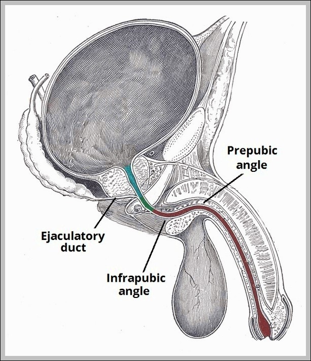
Parts of the Male Urethra. Diagram

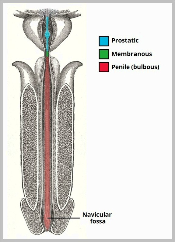
Parts of the Male Urethra Diagram

Male urethra parts: prostatic (through prostate), membranous (shortest, through urogenital diaphragm), spongy/penile (longest, through corpus spongiosum to meatus). It conducts urine/semen.
Borders and Contents of the Adductor Canal. Diagram

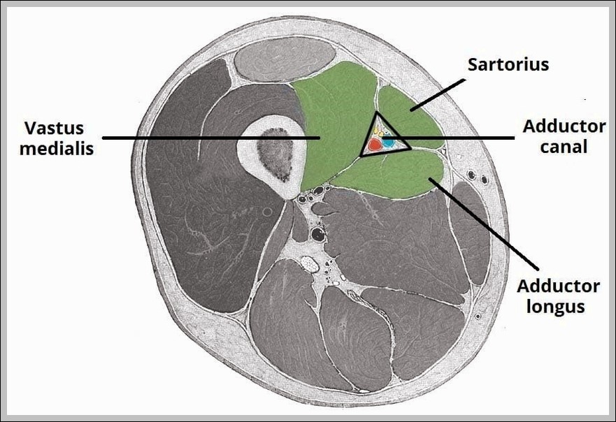
Borders and Contents of the Adductor Canal Diagram

The Borders and Contents of the Adductor Canal diagram defines this subsartorial tunnel from apex of femoral triangle to adductor hiatus: roof (sartorius), anterolateral (vastus medialis), posteromedial (adductors longus/magnus). Contents…
Laparoscopic Cholecystectomy Diagram

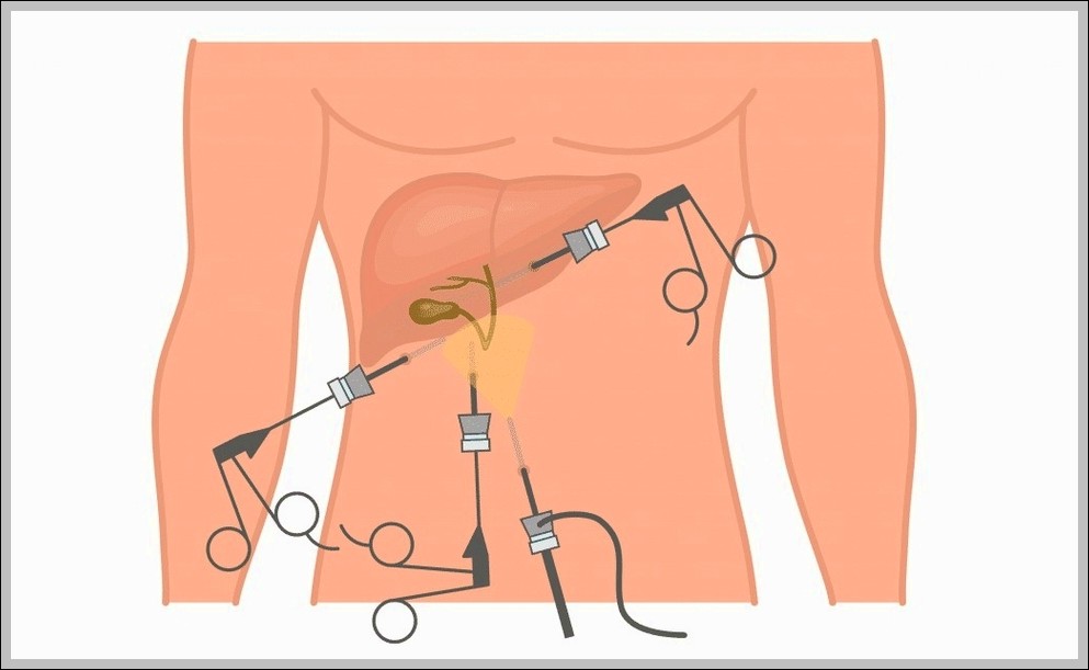
Laparoscopic Cholecystectomy Diagram

This Laparoscopic Cholecystectomy diagram shows the key steps of gallbladder removal using small incisions and a camera. It highlights where the trocars go, the tools in action, and important structures…
Recurrent Branch of Median Nerve Diagram

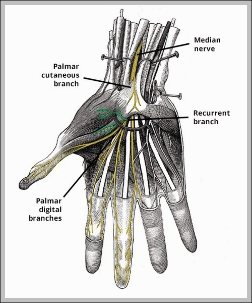
Recurrent Branch of Median Nerve Diagram

Recurrent branch of median nerve arises distal to carpal tunnel, loops around flexor pollicis brevis to supply thenar muscles (abductor pollicis brevis, opponens pollicis, superficial flexor pollicis brevis head). It’s…
Bony Landmarks of the Pubis Diagram

Bony Landmarks of the Pubis Diagram

Pubis bony landmarks: body (anterior), superior/inferior rami, pubic tubercle (inguinal ligament attachment), pubic crest, pectineal line, symphyseal surface.
Internal Structure of the Breast Diagram

Internal Structure of the Breast Diagram

Breast internal structure includes 15-20 lobes of glandular tissue draining via lactiferous ducts to nipple, embedded in fat/connective tissue. Cooper’s ligaments provide support; Cooper suspensory.
Anatomical Position of the Spleen Diagram

Anatomical Position of the Spleen Diagram

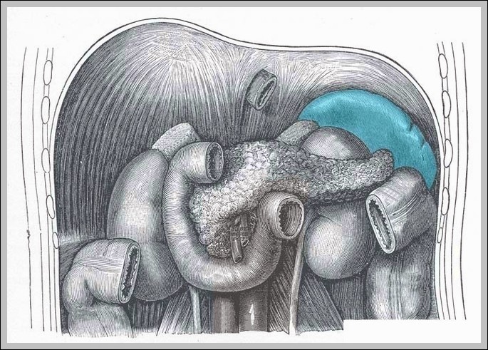
The spleen lies in left upper quadrant, posterior to stomach, protected by ribs 9-11, with diaphragmatic and visceral surfaces, hilum medially for vessels/lymphatics/nerves. It filters blood, stores platelets, and mounts…
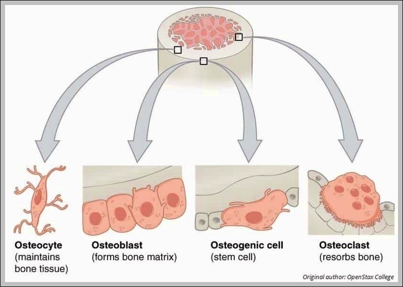
Cellular Components of Bone Diagram

Cellular Components of Bone Diagram

The Cellular Components of Bone diagram zooms to the microscopic level with osteoblasts building matrix, osteocytes living inside lacunae, osteoclasts breaking bone down, and the osteon units with their central…