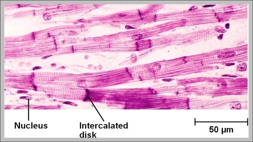
Cardiac Muscle Fibers Image

Cardiac muscle, also known as heart muscle, is the layer of muscle tissue which lies between the endocardium and epicardium. These inner and outer layers of the heart, respectively, surround the cardiac muscle tissue and separate it from the blood and other organs. Cardiac muscle is made from sheets of cardiac muscle cells.
The cardiac muscle fibers are shorter than the skeletal muscle fibers and show the branching pattern. These muscle fibers have one or two centrally placed nuclei. From the cardiac muscle histology slide, you might identify the following important histological features.

Cardiac Muscle Fibers Image Diagram - Cardiac Muscle Fibers Image Chart - Human anatomy diagrams and charts explained. This anatomy system diagram depicts Cardiac Muscle Fibers Image with parts and labels. Best diagram to help learn about health, human body and medicine.

Cardiac Muscle Fibers Image