


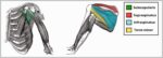


Posted inMedical
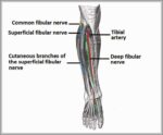
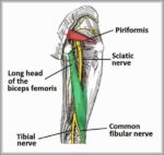
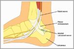
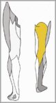
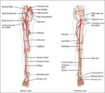
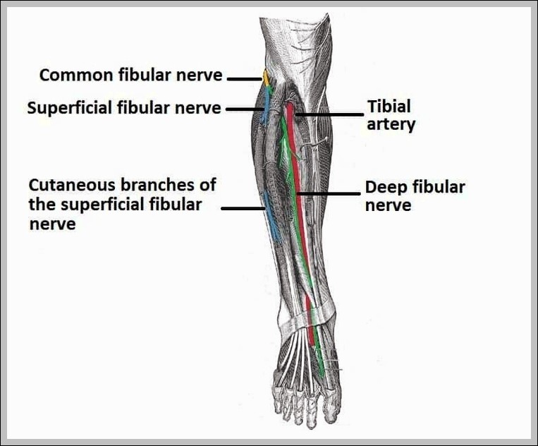
Anatomical Course of the Common Fibular Nerve and its Terminal Branches Diagram
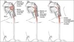
The common fibular (peroneal) nerve wraps around fibular neck (vulnerable to injury), divides into superficial (lateral compartment, peroneus longus/brevis) and deep (anterior compartment, tibialis anterior, extensors) branches. It supplies dorsiflexion,…