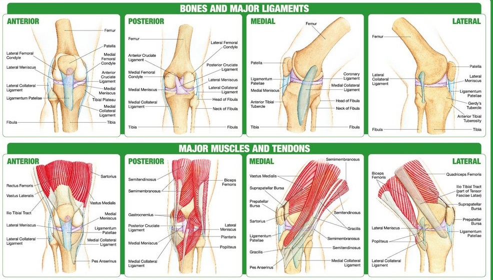
Knee joint anatomy involves looking at each of the different structures in and around the knee. The knee joint is the largest and one of the most complex joints in the human body.
Picture of the Knee. The knee is one of the largest and most complex joints in the body. The knee joins the thigh bone (femur) to the shin bone (tibia). The smaller bone that runs alongside the tibia (fibula) and the kneecap (patella) are the other bones that make the knee joint.
Our knee muscles are responsible for initiating and controlling movement of the knee and the kneecap. They also work with the various buttock, thigh and calf muscles to help control the hip and foot.