The first cervical vertebrae (C1) bears the name of Atlas who was a Titan in Greek mythology.
The lower section consists of the t hird cervical vertebrae (C3) through seventh cervical vertebrae (C7). These spinal bones attach to the thoracic spine and work together to support the head. The fourth cervical vertebra (C4) is centrally located in the cervical (or neck) region of the spinal column.
The lower section consists of the t hird cervical vertebrae (C3) through seventh cervical vertebrae (C7). These spinal bones attach to the thoracic spine and work together to support the head. The fourth cervical vertebra (C4) is centrally located in the cervical (or neck) region of the spinal column.

First Cervical Vertebra Image
Posted inDiagrams
First Cervical Vertebra Image
Post navigation
Previous Post
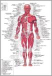 Human Anatomy And Physiology Diagrams Image
Next Post
Knee Muscle Image