Thoracic Vertebra Image

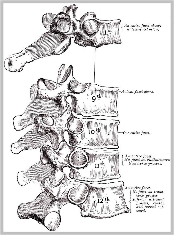
This article will elucidate all the mysteries surrounding the thoracic vertebrae and will describe both their typical and atypical features. The thoracic vertebrae are located in the middle section of the vertebral column, specifically inferior to the cervical vertebrae and superior to the lumbar vertebrae.
T1 (1st Thoracic Vertebra) The T1 vertebra is the first (uppermost) of the twelve (12) thoracic vertebrae that make up the central and largest section of the spinal column between the lumbar vertebrae below and the cervical vertebrae above. While larger than the C7 vertebra above it, the T1 is the smallest of the thoracic vertebrae.

Thoracic Vertebra Image Diagram - Thoracic Vertebra Image Chart - Human anatomy diagrams and charts explained. This anatomy system diagram depicts Thoracic Vertebra Image with parts and labels. Best diagram to help learn about health, human body and medicine.

Thoracic Vertebra Image

First Cervical Vertebra Image

The first cervical vertebrae (C1) bears the name of Atlas who was a Titan in Greek mythology.
The lower section consists of the t hird cervical vertebrae (C3) through seventh cervical vertebrae (C7). These spinal bones attach to the thoracic spine and work together to support the head. The fourth cervical vertebra (C4) is centrally located in the cervical (or neck) region of the spinal column.

First Cervical Vertebra Image Diagram - First Cervical Vertebra Image Chart - Human anatomy diagrams and charts explained. This anatomy system diagram depicts First Cervical Vertebra Image with parts and labels. Best diagram to help learn about health, human body and medicine.

First Cervical Vertebra Image